Copyright
©The Author(s) 2023.
World J Clin Cases. Oct 16, 2023; 11(29): 7156-7161
Published online Oct 16, 2023. doi: 10.12998/wjcc.v11.i29.7156
Published online Oct 16, 2023. doi: 10.12998/wjcc.v11.i29.7156
Figure 1 Bone marrow biopsy after admission suggested acute myelocytic leukemia.
The relatively active proliferation of bone marrow nucleated cells and the increased ratio of granuloid/erythroid cells. The granuloids are dominated by the cells that are biased towards the naive stage. Immunohistochemistry shows the following: CD34 small blood vessels (+), a large quantity of prokaryotic cells (+), a large quantity of CD117 (+), a large quantity of MPO (+), a large quantity of CD99 (+), TdT (-), a small quantity of CD3 (+), CD79a (-), and lysozyme sporadic (+).
Figure 2 Bone marrow smear shows a myelogram of acute myelocytic leukemia inclined to M2 type.
Blood smear: Number of leukocytes is on the large side, and the number of primitive cells accounts for 74%; mature erythrocytes vary in size; a small number of sporadic platelets are visible. Bone marrow smear: Myelogram of acute myelocytic leukemia is inclined to M2 type.
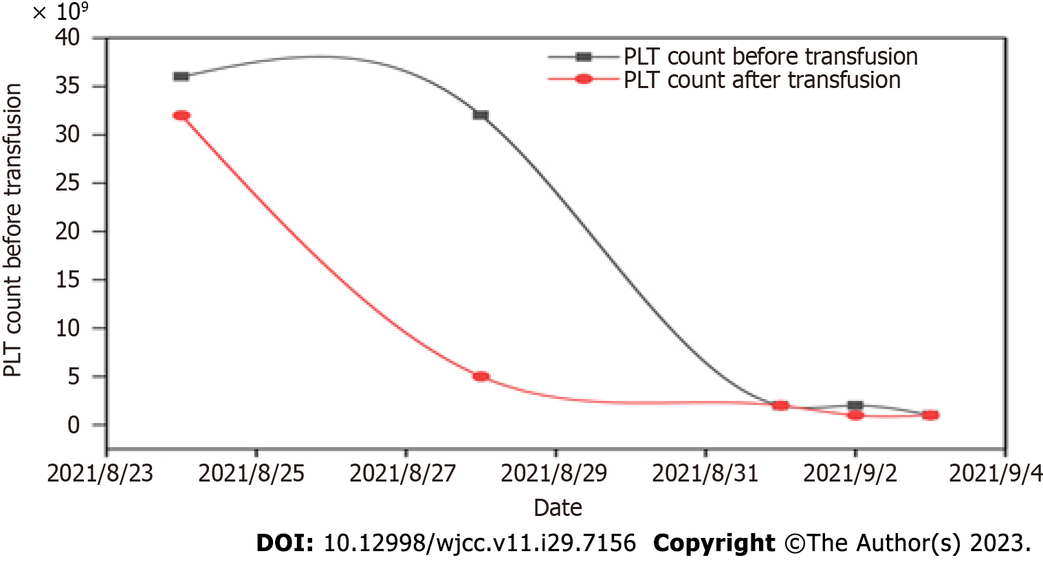
Figure 3 The efficacy of platelet transfusion after myelosuppression caused by the first "IA" chemotherapy.
PLT: Platelet.
Figure 4 The efficacy of platelet transfusion after myelosuppression caused by the second cycle of "IA" chemotherapy.
PLT: Platelet.
- Citation: Tu SK, Fan HJ, Shi ZW, Li XL, Li M, Song K. First platelet transfusion refractoriness in a patient with acute myelocytic leukemia: A case report. World J Clin Cases 2023; 11(29): 7156-7161
- URL: https://www.wjgnet.com/2307-8960/full/v11/i29/7156.htm
- DOI: https://dx.doi.org/10.12998/wjcc.v11.i29.7156


