Copyright
©The Author(s) 2023.
World J Clin Cases. Oct 16, 2023; 11(29): 6995-7003
Published online Oct 16, 2023. doi: 10.12998/wjcc.v11.i29.6995
Published online Oct 16, 2023. doi: 10.12998/wjcc.v11.i29.6995
Figure 1 Colorectal sessile serrated lesions characterized by colonoscopy demonstrate.
A: White light endoscopy of the sessile serrated lesion (SSL) surface covered with mucus cap; B: White light endoscopy of the SSL with pale color and cloud-like surface; C: White light endoscopy of the SSL-dysplasia (SSL-D) against a pale color background with localized reddish mucosa and a depression visible in the center of the cloud-like surface; D: In the endoscopic blue laser imaging (BLI) mode, the dilated crypt of the SSL appears as black spots (as shown by the arrow); E: In the endoscopic magnified BLI mode, microvascular varicose is visible on the surface of the SSL-D (as shown by arrows); F: In endoscopic magnified BLI mode, the dilated crypt of the SSL-D appears as Pit IIIL type in the background of Pit II-O.
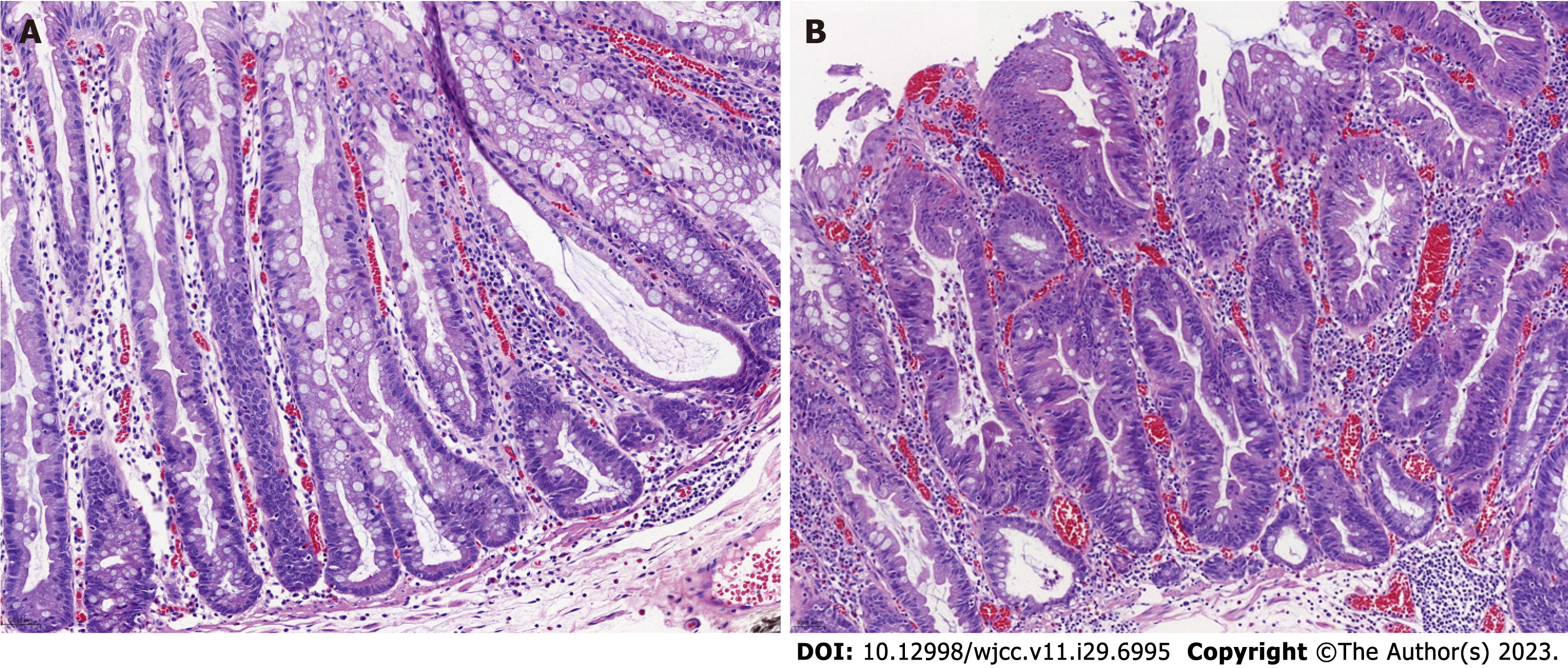
Figure 2 Histopathological demonstration of colorectal the sessile serrated lesion and sessile serrated lesion-dysplasia (hematoxylin and eosin, objective magnifications 20 ×, 3DHISTECH).
A: The crypt has at least one of the following histologic features: (1) Horizontal growth along the mucosal muscle layer (L or inverted T-shaped crypt structure); (2) Expansion of the crypt base (basal 1/3 of the crypt base); (3) Jagged swelling of the crypt base; and (4) Asymmetric hyperplasia (proliferative band extending laterally from the base); B: Complex structural abnormalities, including: (1) Crypt elongation, crowding, complex branching, sieve-like structures, and villi-like structures; and (2) Cytologic abnormalities of diverse morphology, either with cuboidal cells, eosinophilic cytoplasm, vesicular nuclei, and prominent nucleoli, or with elongated cells, eosinophilic cytoplasm, deep-stained nuclei, and pseudostratified nuclei, with common nuclear schizophrenic signs.
- Citation: Wang RG, Ren YT, Jiang X, Wei L, Zhang XF, Liu H, Jiang B. Usefulness of analyzing endoscopic features in identifying the colorectal serrated sessile lesions with and without dysplasia. World J Clin Cases 2023; 11(29): 6995-7003
- URL: https://www.wjgnet.com/2307-8960/full/v11/i29/6995.htm
- DOI: https://dx.doi.org/10.12998/wjcc.v11.i29.6995


