Copyright
©The Author(s) 2022.
World J Clin Cases. Dec 16, 2022; 10(35): 13052-13057
Published online Dec 16, 2022. doi: 10.12998/wjcc.v10.i35.13052
Published online Dec 16, 2022. doi: 10.12998/wjcc.v10.i35.13052
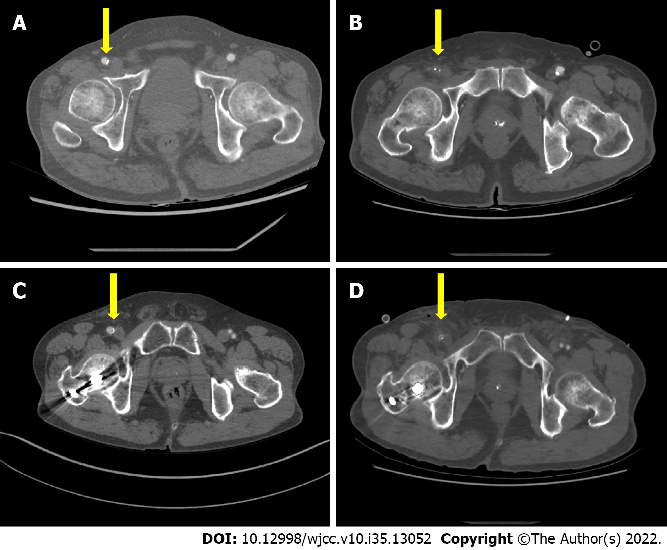
Figure 1 Comparison of computed tomography findings before and after surgery.
A: Computed tomography (CT) performed before minimally invasive cardiac surgery in case 1. The yellow arrow indicates the right common femoral artery, and although calcification is present, no significant stenosis is observed; B: CT performed after minimally invasive cardiac surgery in case 1. The yellow arrow indicates the right common femoral artery, and it can be seen that the inside of the right common femoral artery is not contrasted. C: CT performed prior to minimally invasive cardiac surgery in case 2. Yellow arrow indicates the right common femoral artery. Calcification is observed, but no significant stenosis is observed; D: CT performed after minimally invasive cardiac surgery in case 2. The yellow arrow indicates the right common femoral artery, and it can be seen that the inside of the right common femoral artery is not contrasted.
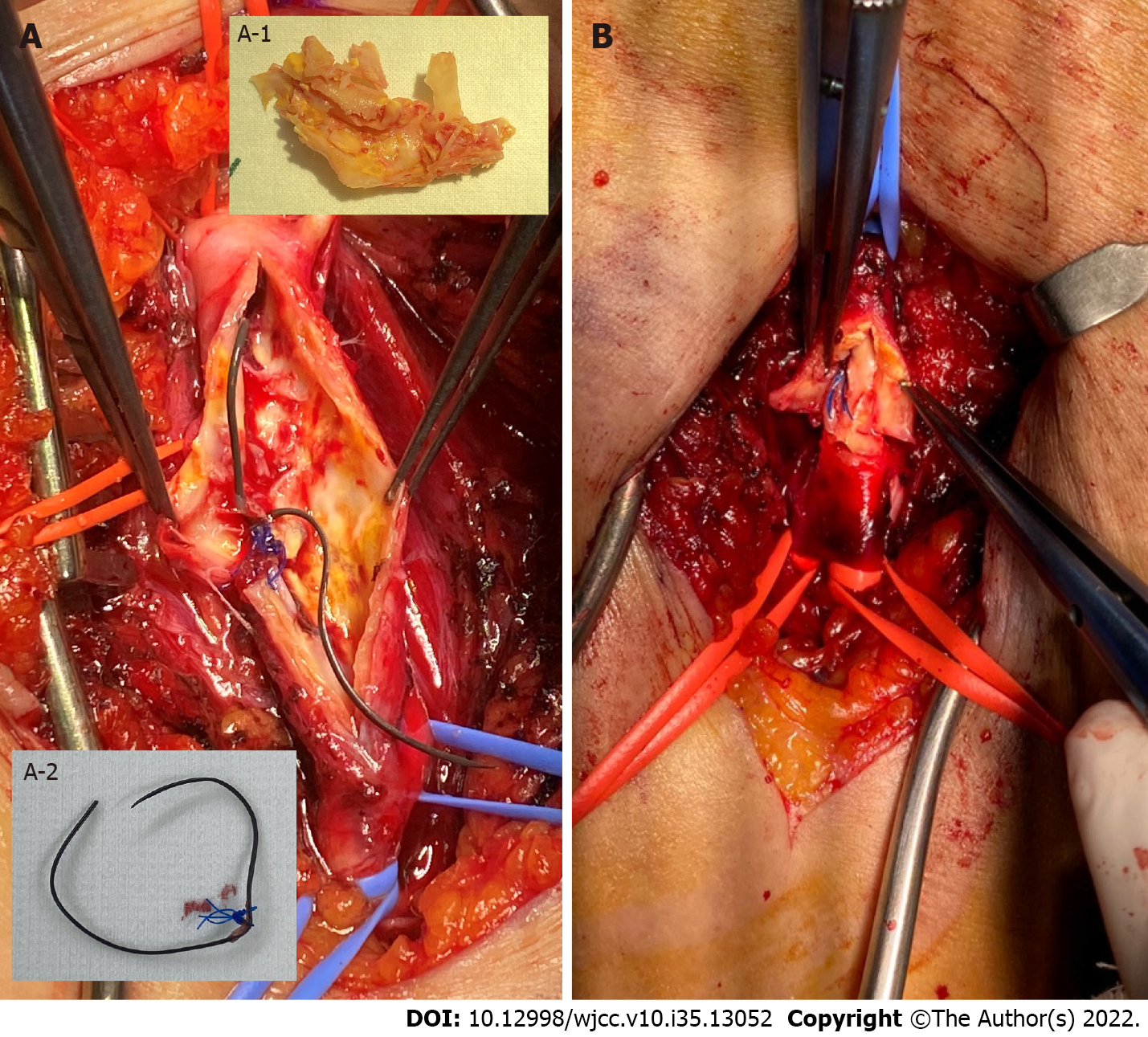
Figure 2 Right common femoral artery.
A: Inside the right common femoral artery in case 1. A thread from the Perclose ProGlide™ SMC System (Abbott, IL, USA) is seen tied over the intima on the dorsal side of the vessel; A-1: Atheroma removed by endarterectomy; A-2: Tie thread of Perclose ProGlide™ SMC System removed from the vessel wall; B: Inside the right common femoral artery in case 2. As in case 1, a thread of the Perclose ProGlide™ SMC system is seen tied on the intima on the dorsal side of the vessel.
- Citation: Lee J, Huh U, Song S, Lee CW. Acute limb ischemia after minimally invasive cardiac surgery using the ProGlide: A case series. World J Clin Cases 2022; 10(35): 13052-13057
- URL: https://www.wjgnet.com/2307-8960/full/v10/i35/13052.htm
- DOI: https://dx.doi.org/10.12998/wjcc.v10.i35.13052


