Copyright
©The Author(s) 2022.
World J Clin Cases. Nov 26, 2022; 10(33): 12404-12409
Published online Nov 26, 2022. doi: 10.12998/wjcc.v10.i33.12404
Published online Nov 26, 2022. doi: 10.12998/wjcc.v10.i33.12404
Figure 1 Computed tomography findings.
A: Computed tomography (CT) imaging findings for the two left enlarged lateral lymph nodes; B: CT imaging findings for the right enlarged lateral lymph node; C: CT imaging findings for the largest mesorectal lymph node (arrow).
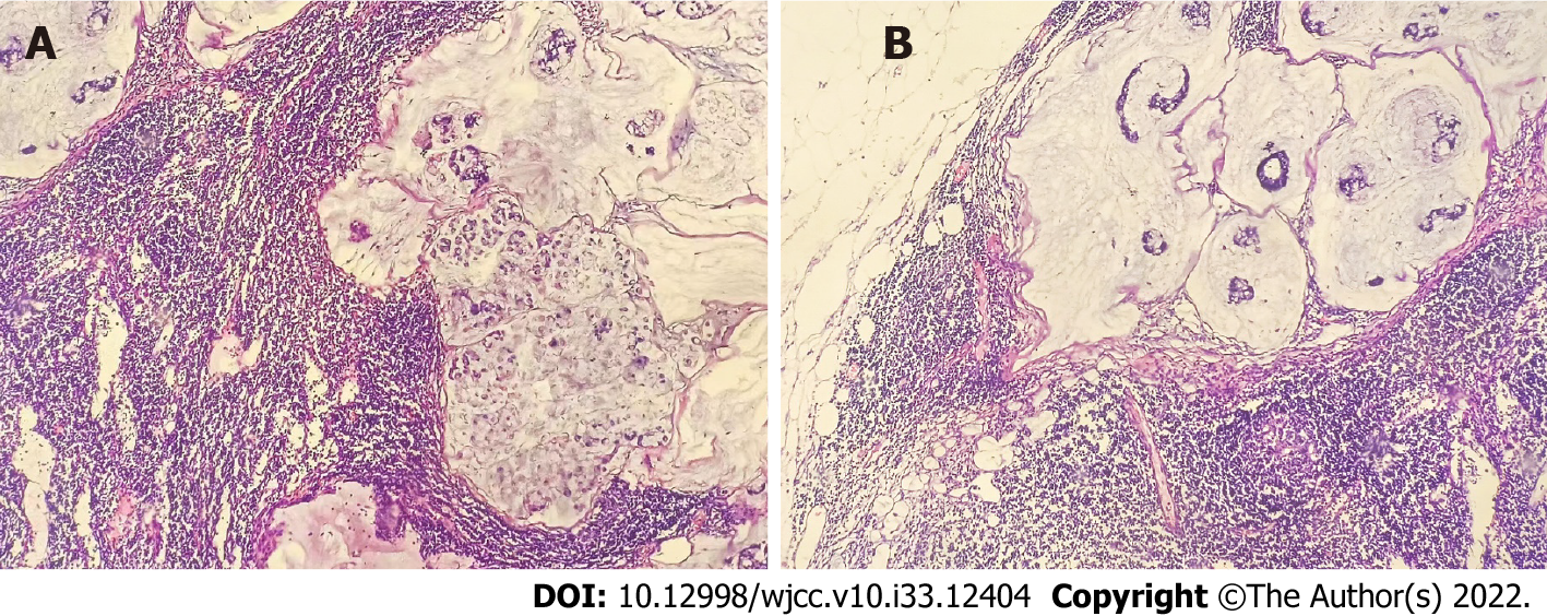
Figure 2 Pathological imaging for the two positive left lateral lymph nodes (hematoxylin and eosin × 100).
A and B: Pathological images of two positive lateral lymph nodes, respectively. The lymphatic structure is destroyed, and numerous mucous lakes are formed in which floating adenosine cells are observed.
- Citation: Liu XW, Zhou B, Wu XY, Yu WB, Zhu RF. T1 rectal mucinous adenocarcinoma with bilateral enlarged lateral lymph nodes and unilateral metastasis: A case report. World J Clin Cases 2022; 10(33): 12404-12409
- URL: https://www.wjgnet.com/2307-8960/full/v10/i33/12404.htm
- DOI: https://dx.doi.org/10.12998/wjcc.v10.i33.12404


