Copyright
©The Author(s) 2022.
World J Clin Cases. Nov 26, 2022; 10(33): 12365-12374
Published online Nov 26, 2022. doi: 10.12998/wjcc.v10.i33.12365
Published online Nov 26, 2022. doi: 10.12998/wjcc.v10.i33.12365
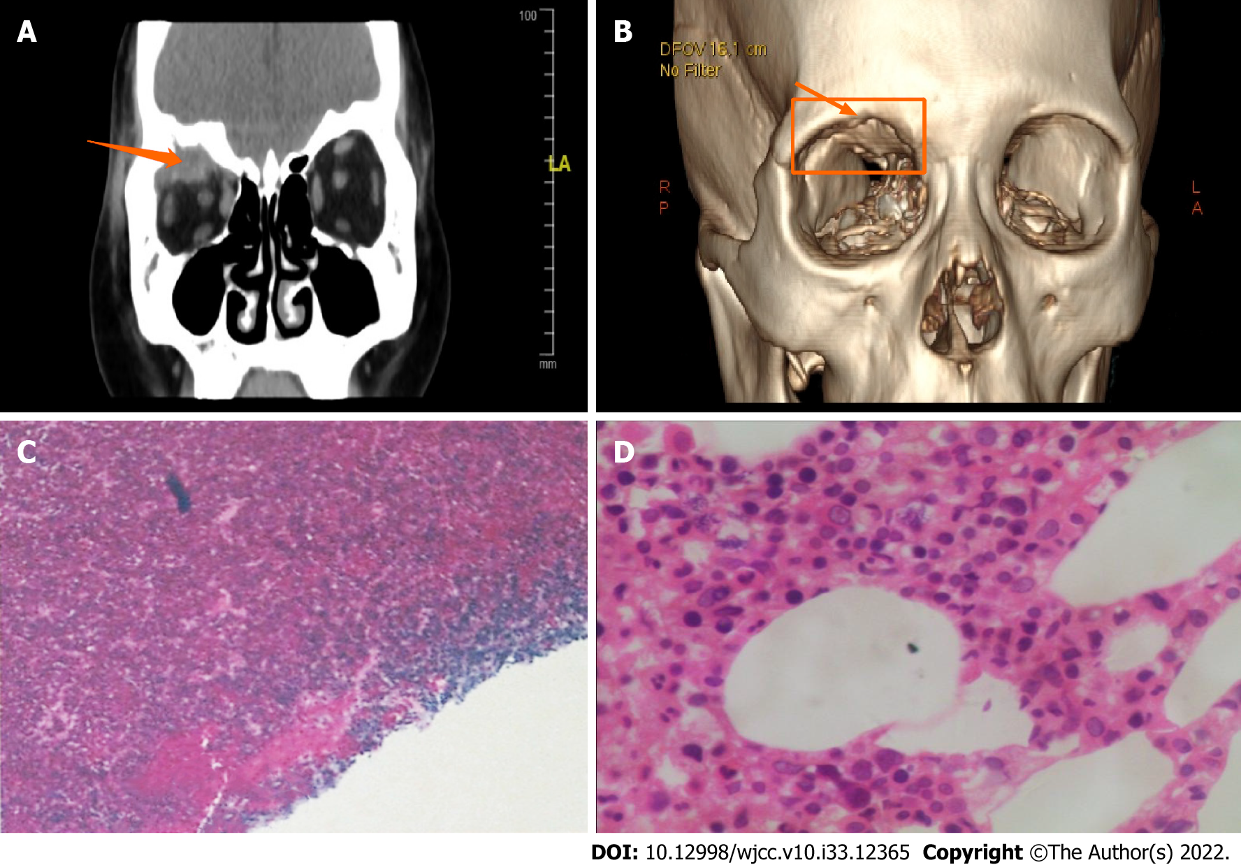
Figure 1 Computed tomography image and histology.
A: Orbital extramedullary disease (EMD) in computed tomography (CT) image; B: Three D reconstruction of CT images of orbital EMD; C: Pathological findings of biopsy tissue from an orbital mass. It is showed in immunohistochemistry (HE stain 100×, size approx. 4 cm × 3 cm × 1 cm): CD10(-), CD138(+), CD15(-), CD19(-), CD20(-), CD3(-), CD34(-), CD38(+), CD56 (scattered +), CD99(+), CK(-), EMA(+), IgG(+), Ki-67 index (30%), Mpo(-), MUM-1(+), TdT(-), k(+), λ(-), CD79α(-), EBER(-). The diagnosis of plasma cell tumor is considered based on the medical history and immunohistochemical findings; D: Bone marrow images while MM with EMD occurred. The proportion of naive plasma cells is 0.5% in HE staining (400×).
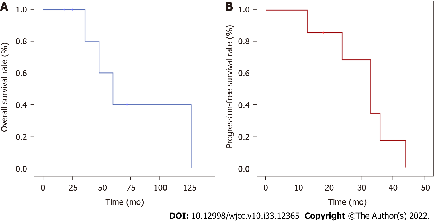
Figure 2 Impact of orbital extramedullary disease on the survival.
A: Impact of orbital extramedullary disease on overall survival; B: Impact of orbital extramedullary disease on progression free survival.
- Citation: Hu WL, Song JY, Li X, Pei XJ, Zhang JJ, Shen M, Tang R, Pan ZY, Huang ZX. Clinical features and prognosis of multiple myeloma and orbital extramedullary disease: Seven cases report and review of literature. World J Clin Cases 2022; 10(33): 12365-12374
- URL: https://www.wjgnet.com/2307-8960/full/v10/i33/12365.htm
- DOI: https://dx.doi.org/10.12998/wjcc.v10.i33.12365


