Copyright
©The Author(s) 2022.
World J Clin Cases. Nov 26, 2022; 10(33): 12352-12357
Published online Nov 26, 2022. doi: 10.12998/wjcc.v10.i33.12352
Published online Nov 26, 2022. doi: 10.12998/wjcc.v10.i33.12352
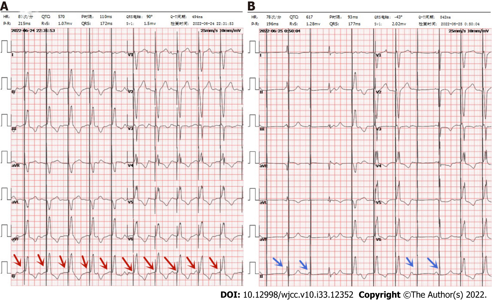
Figure 1 Electrocardiogram.
A: Electrocardiogram (ECG) in emergency department: red arrow indicates normal pacing; B: ECG during amaurosis: Blue arrows indicate poor pacing.
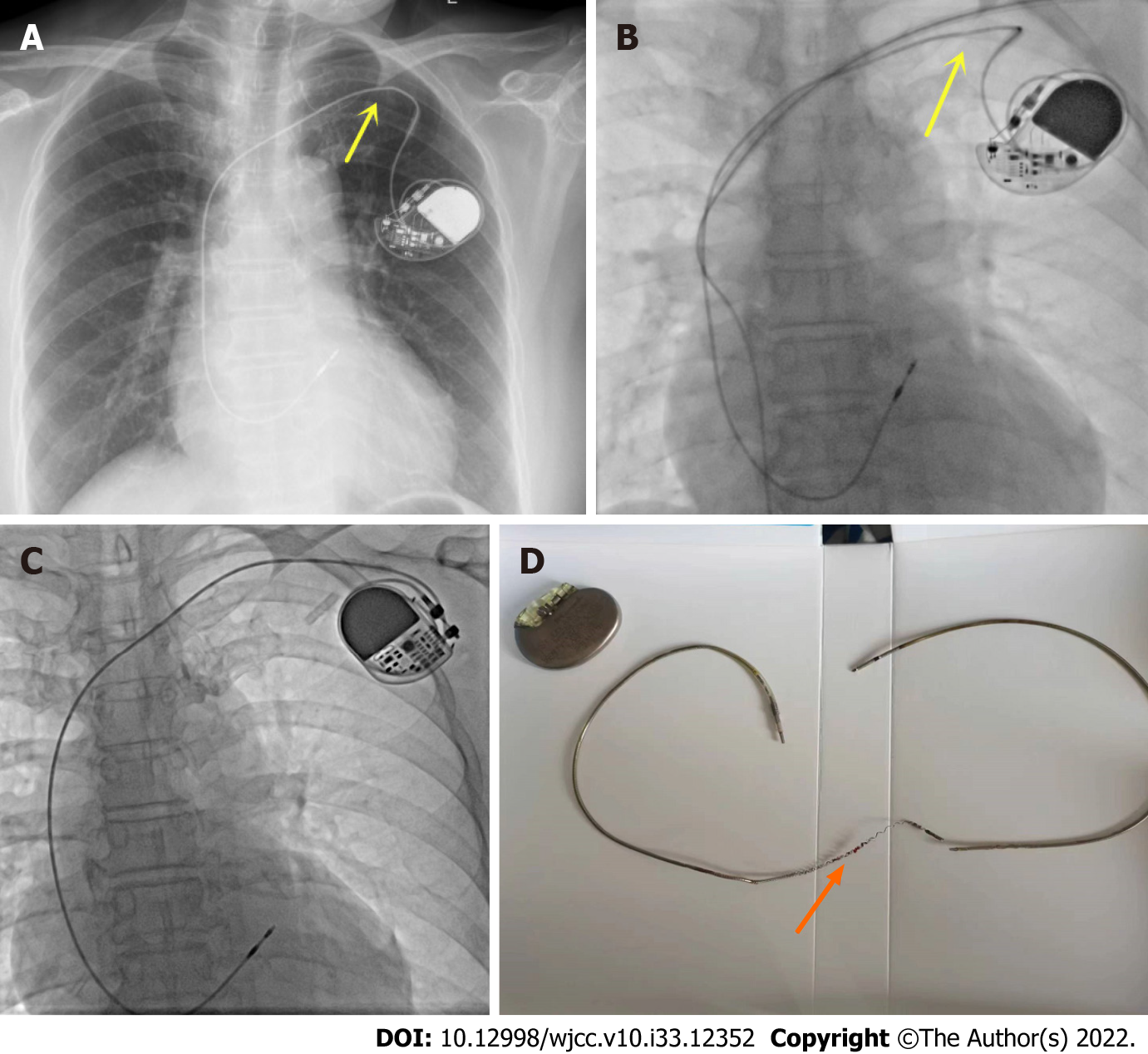
Figure 2 Brain computed tomography/electrodes and pacemaker.
A and B: The fluoroscopic image shows the lead depression in the subclavian area (yellow arrows); C: Replaced pacemaker and pacemaker electrode; D: Extracted lead shows broken lead (orange arrow).
- Citation: Zhu XY, Tang XH, Huang WY. Pacemaker electrode rupture causes recurrent syncope: A case report. World J Clin Cases 2022; 10(33): 12352-12357
- URL: https://www.wjgnet.com/2307-8960/full/v10/i33/12352.htm
- DOI: https://dx.doi.org/10.12998/wjcc.v10.i33.12352


