Copyright
©The Author(s) 2022.
World J Clin Cases. Nov 26, 2022; 10(33): 12268-12277
Published online Nov 26, 2022. doi: 10.12998/wjcc.v10.i33.12268
Published online Nov 26, 2022. doi: 10.12998/wjcc.v10.i33.12268
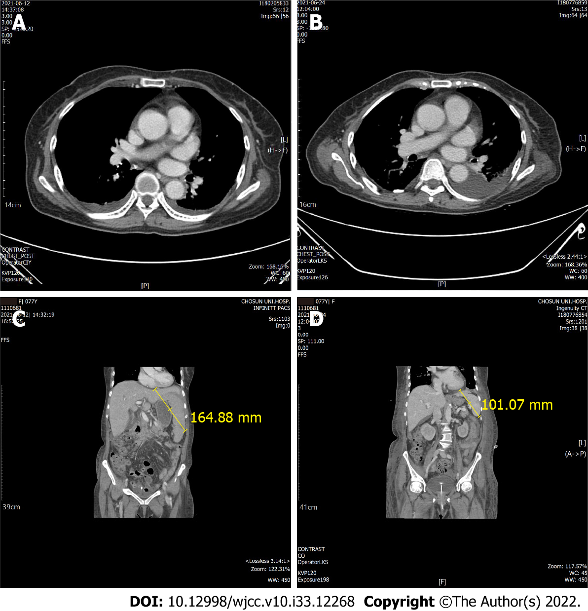
Figure 1 Computed tomography imaging.
A: Initial thorax imaging; B: Day 13 thorax imaging; C: Initial hospitalization; D: Day 13 of hospitalization. Thorax computed tomography showed no findings indicating infection, but splenomegaly and liver cirrhosis were confirmed on abdomino-pelvic computed tomography. Splenomegaly improved on day 13 of hospitalization.
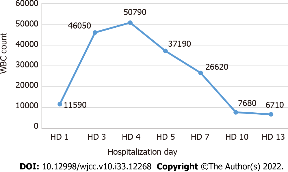
Figure 2 White blood cell count during hospitalization.
WBC: White blood cell; HD: Hospitalization day.
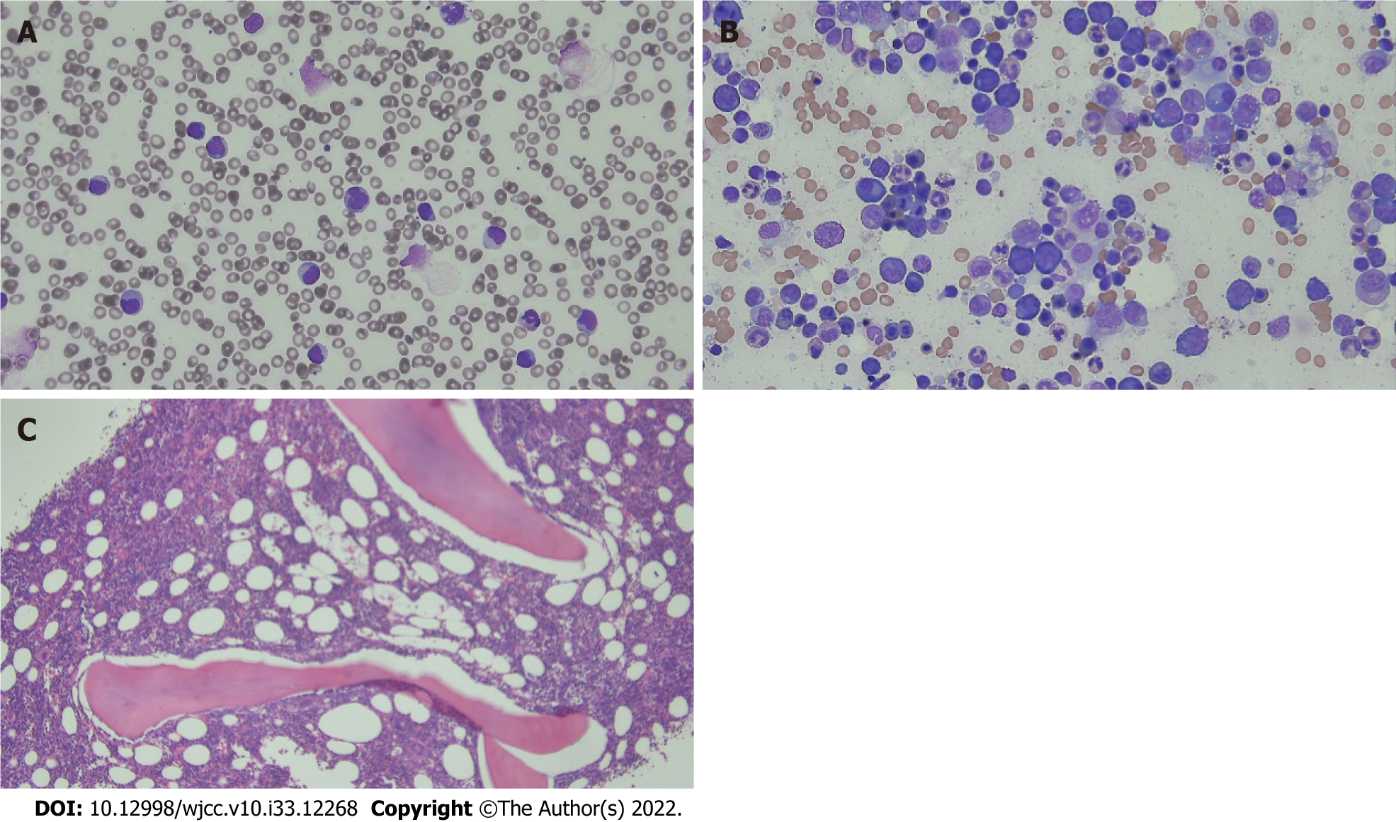
Figure 3 Peripheral blood smear and bone marrow examination.
A: Peripheral blood smear; B: Bone marrow aspiration; C: Bone marrow biopsy. Peripheral blood smear showed leukocytosis with neutrophils and immature cells. Bone marrow aspiration and biopsy sample revealed reactive marrow.
- Citation: Lee SB, Park CY, Park SG, Lee HJ. Case mistaken for leukemia after mRNA COVID-19 vaccine administration: A case report. World J Clin Cases 2022; 10(33): 12268-12277
- URL: https://www.wjgnet.com/2307-8960/full/v10/i33/12268.htm
- DOI: https://dx.doi.org/10.12998/wjcc.v10.i33.12268


