Copyright
©The Author(s) 2022.
World J Clin Cases. Nov 6, 2022; 10(31): 11607-11616
Published online Nov 6, 2022. doi: 10.12998/wjcc.v10.i31.11607
Published online Nov 6, 2022. doi: 10.12998/wjcc.v10.i31.11607
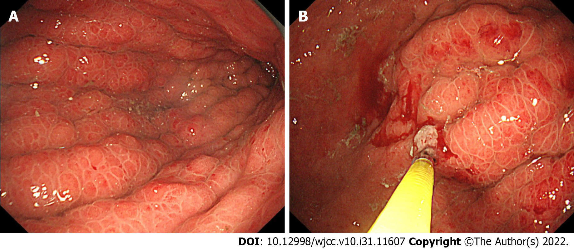
Figure 1 An endoscopic examination of gastric linitis plastica.
A: Endoscopic findings showed giant gastric folds and reddish mucosa; however, no epithelial changes were observed. The gastric lumen was not distensible by air inflation; B: Seven specimens were obtained from the gastric mucosa, none of which showed malignancy.
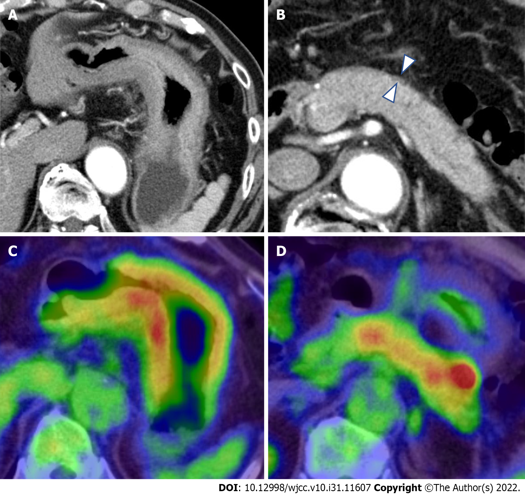
Figure 2 Computed tomography and 18F-Fluorodeoxyglucose-positron emission tomography/computed tomography findings.
A: Contrast-enhanced computed tomography (CT) showed thickening of the wall of the gastric body; B: CT showed the diffuse enlargement of the pancreas and peripancreatic rim (arrowheads); C and D: Fluorodeoxyglucose-positron emission tomography/CT (FDG-PET/CT) showed the accumulation of FDG within both the gastric wall (SUVmax: 19.2) and pancreas.
Figure 3 Endoscopic ultrasonography and endoscopic ultrasonography-guided fine-needle biopsy findings.
A: Under endoscopic ultrasonography (EUS), the third layer representing the submucosa (arrowhead) and the fourth layer representing the muscularis propria (arrow) were thickened; B: An EUS-fine-needle biops (FNB) of the thickened gastric wall was performed with a 19-gauge needle; C: EUS revealed hyperechoic spots in the diffuse hypoechoic pancreatic parenchymal and duct-penetrating sign (red arrow).
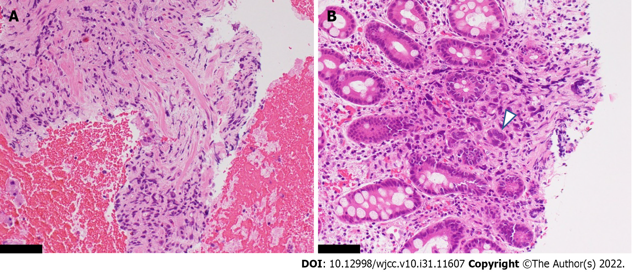
Figure 4 Histopathological specimen obtained by an endoscopic ultrasonography-guided fine-needle biopsy of the thickened gastric wall.
A: Within the intricate muscularis propria, fibroblasts were proliferating, while a few scattered cells suspected of malignancy were seen; B: Poorly differentiated adenocarcinoma cells were seen within the deeper portion of the hyperplastic mucosa (arrowhead). The black scale bar represents 250 μm.
Figure 5 Endoscopic and computed tomography findings after the start of chemotherapy.
A and B: After the start of chemotherapy, the endoscopic findings, such as the giant folds, were improved, and the gastric lumen became distensible, which allowed for duodenoscopy; C: Chemotherapy improved the computed tomography findings of the thickened gastric wall and diffuse enlargement of the pancreas.
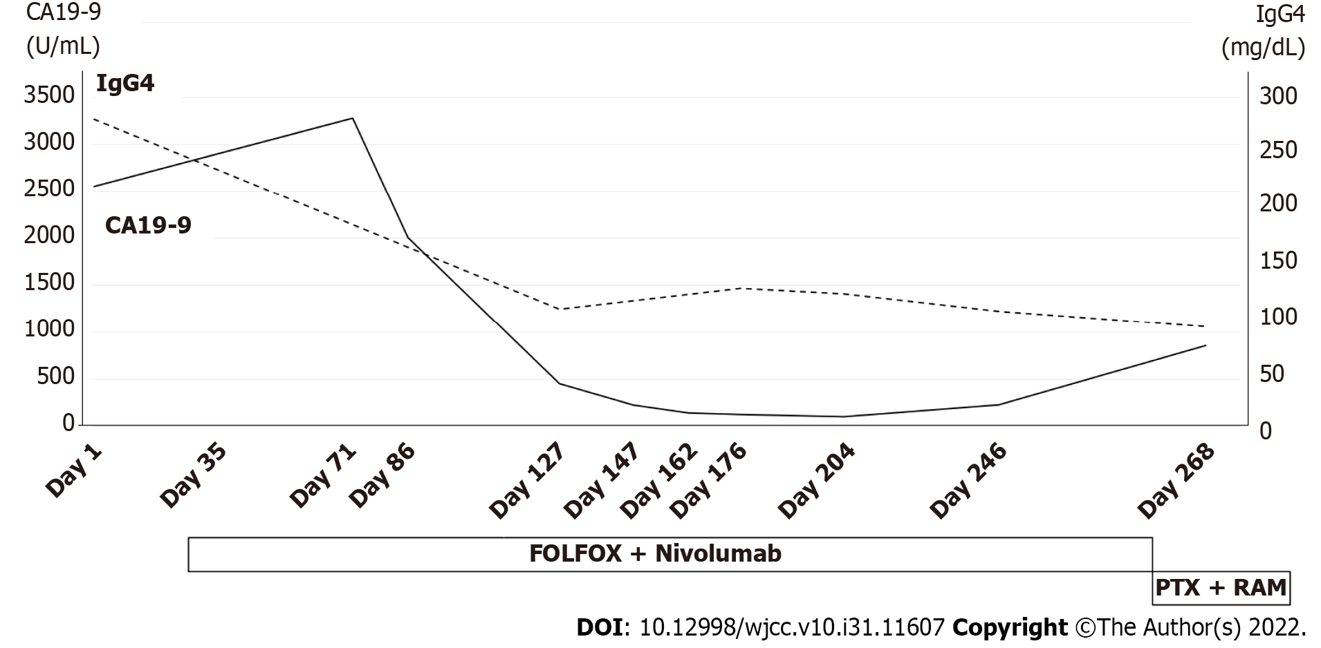
Figure 6 Serum levels of both carbohydrate antigen 19-9 and immunoglobulin-G4 improved during chemotherapy.
CA19-9: Carbohydrate antigen 19-9; IgG4: Immunoglobulin-G4; FOLFOX: 5-fluorouracil, leucovorin and oxaliplatin; PTX: Paclitaxel; RAM: Ramucirumab.
- Citation: Sato R, Matsumoto K, Kanzaki H, Matsumi A, Miyamoto K, Morimoto K, Terasawa H, Fujii Y, Yamazaki T, Uchida D, Tsutsumi K, Horiguchi S, Kato H. Gastric linitis plastica with autoimmune pancreatitis diagnosed by an endoscopic ultrasonography-guided fine-needle biopsy: A case report. World J Clin Cases 2022; 10(31): 11607-11616
- URL: https://www.wjgnet.com/2307-8960/full/v10/i31/11607.htm
- DOI: https://dx.doi.org/10.12998/wjcc.v10.i31.11607


