Copyright
©The Author(s) 2022.
World J Clin Cases. Nov 6, 2022; 10(31): 11555-11560
Published online Nov 6, 2022. doi: 10.12998/wjcc.v10.i31.11555
Published online Nov 6, 2022. doi: 10.12998/wjcc.v10.i31.11555
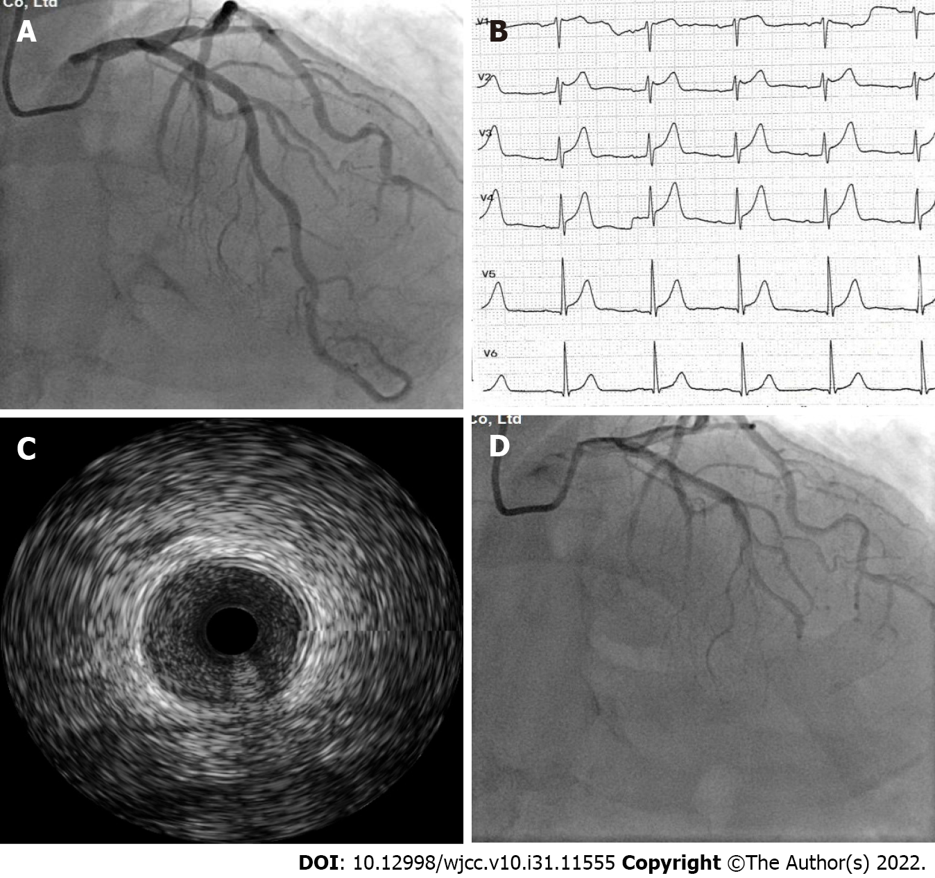
Figure 1 Imaging examination of the patient with Kounis syndrome.
A: Normal left coronary artery angiogram; B: Electrocardiogram during chest pain demonstrated a 2 mm ST-segment elevation in V1-V4 Leads; C: Angiogram revealed left coronary artery vasospasm; D: Intravenous ultrasound image of the left coronary artery vasospasm.
- Citation: Xu GZ, Wang G. Acute myocardial infarction due to Kounis syndrome: A case report. World J Clin Cases 2022; 10(31): 11555-11560
- URL: https://www.wjgnet.com/2307-8960/full/v10/i31/11555.htm
- DOI: https://dx.doi.org/10.12998/wjcc.v10.i31.11555


