Copyright
©The Author(s) 2022.
World J Clin Cases. Nov 6, 2022; 10(31): 11536-11541
Published online Nov 6, 2022. doi: 10.12998/wjcc.v10.i31.11536
Published online Nov 6, 2022. doi: 10.12998/wjcc.v10.i31.11536
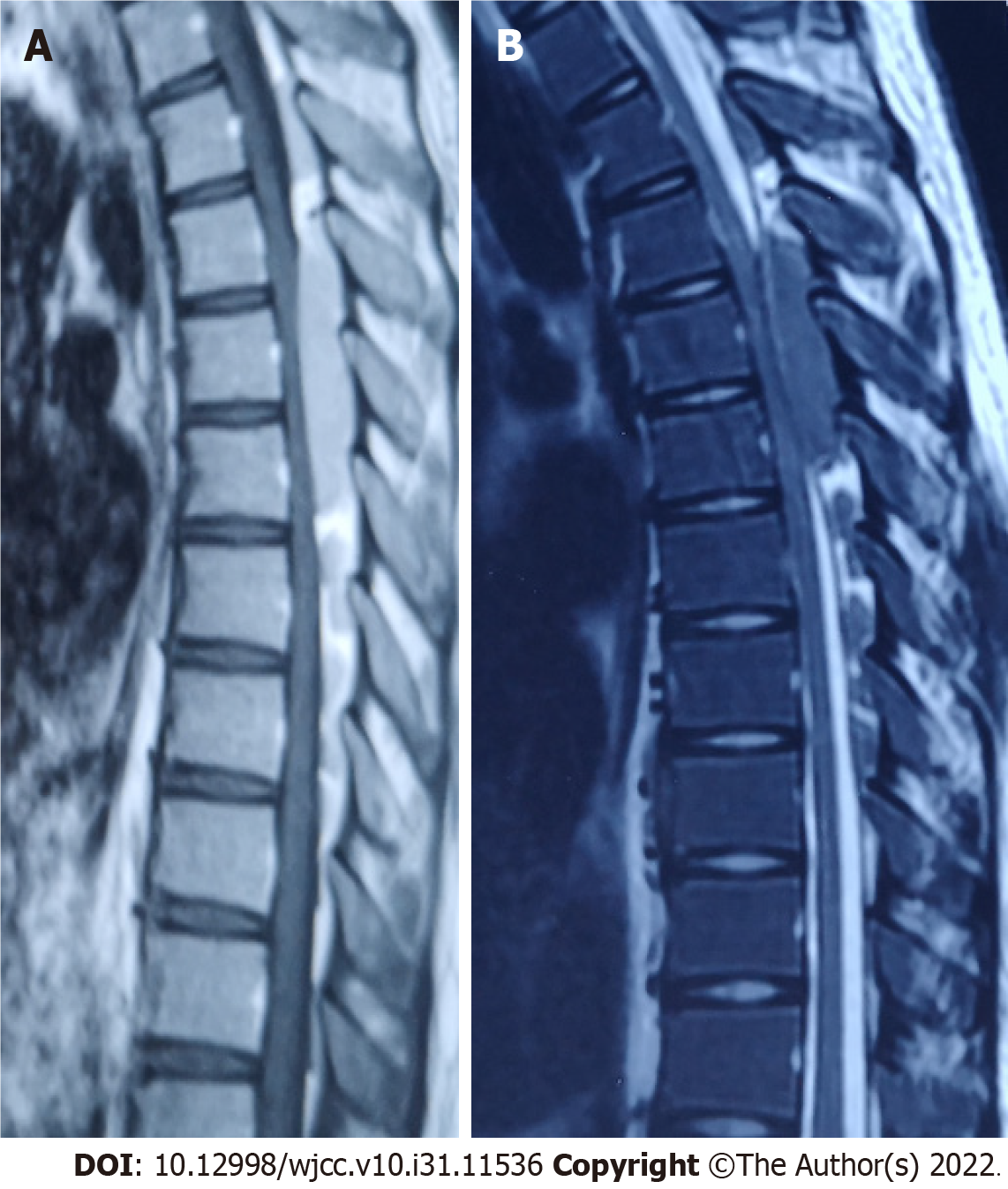
Figure 1 On admission, magnetic resonance imaging of the thoracic spine showed a space-occupying lesion in the long dorsal segment of the T3-8 spinal canal and spinal cord compression.
A: T1-weighted image; B: T2-weighted image.
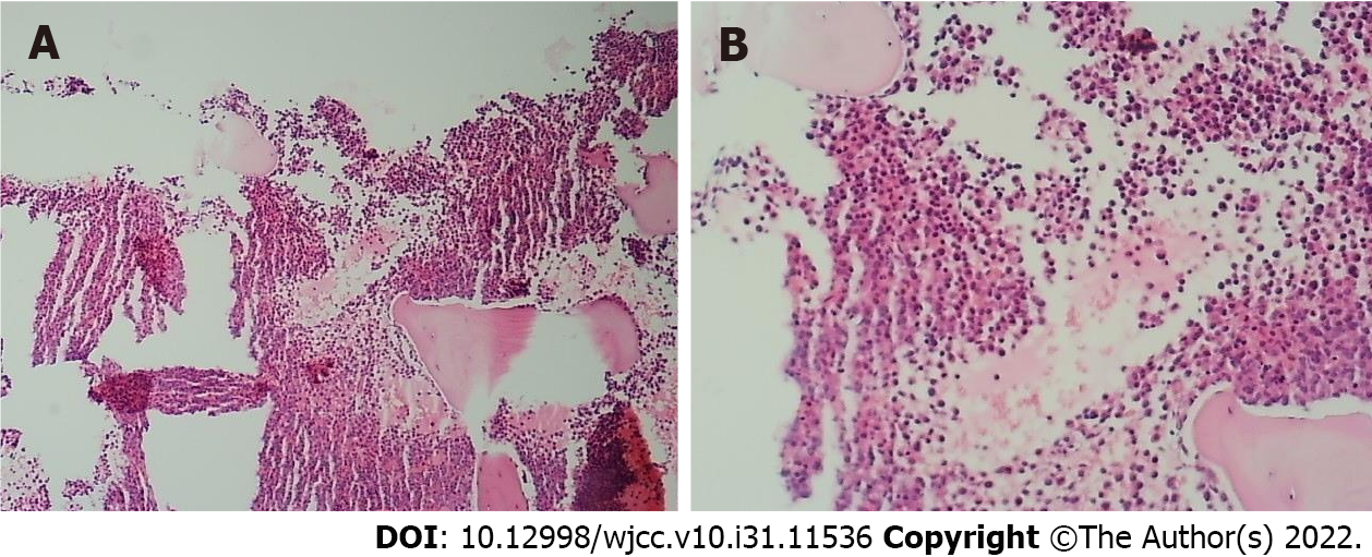
Figure 2 Proliferation of nucleated cells in the bone marrow was extremely active (about 90% of the hematopoietic area).
A: Original magnification, × 40; B: Original magnification, × 400.
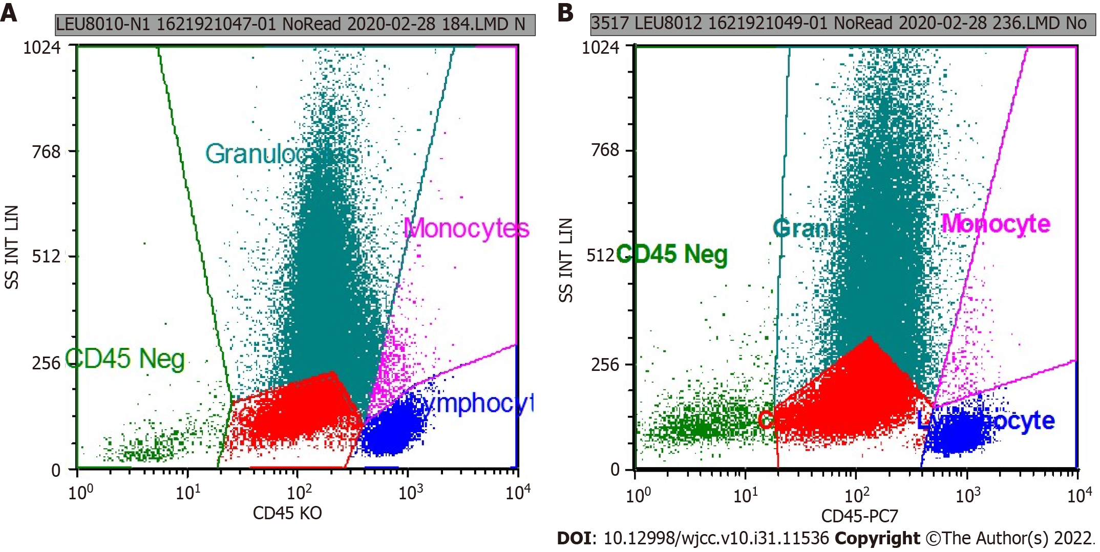
Figure 3 The results of flow cytometry were consistent with the immunophenotype of acute myeloid leukemia (non∙m3).
A: CD117+ cells occupied the total number of nuclear cells (21.87%); B: CD117+ cells occupied the total number of nuclear cells (24.61%).
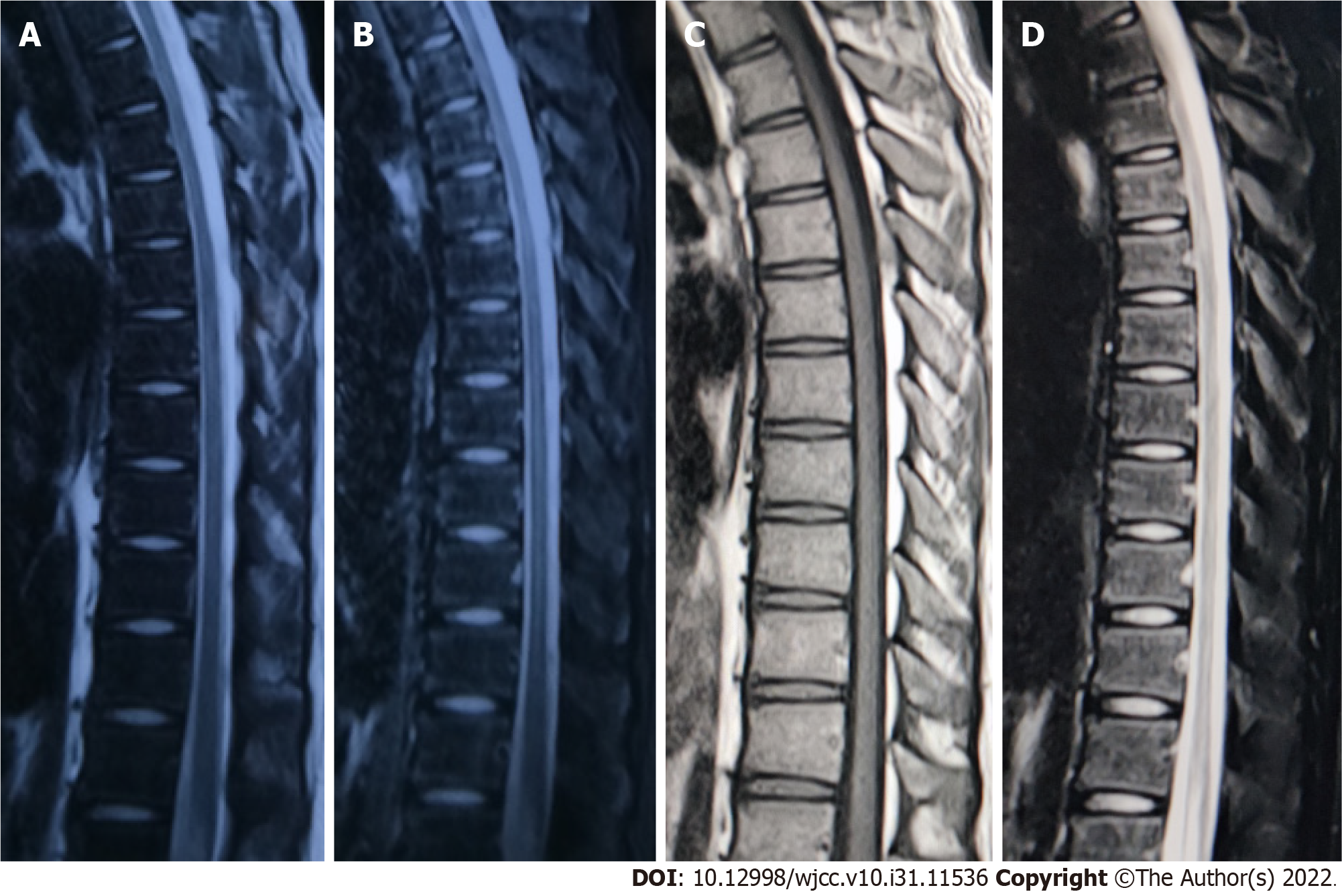
Figure 4 After chemotherapy, magnetic resonance imaging of the thoracic spine was reexamined 1 mo and 1 year later.
The space-occupying lesion in the spinal canal had disappeared completely, and no compression of the spinal cord was observed. A: T2-weighted image; B: T2-weighted image, fat-suppression; C: T1-weighted image; D: T1-weighted image, fat-suppression.
- Citation: Shao YD, Wang XH, Sun L, Cui XG. Granulocytic sarcoma with long spinal cord compression: A case report. World J Clin Cases 2022; 10(31): 11536-11541
- URL: https://www.wjgnet.com/2307-8960/full/v10/i31/11536.htm
- DOI: https://dx.doi.org/10.12998/wjcc.v10.i31.11536


