Copyright
©The Author(s) 2022.
World J Clin Cases. Oct 26, 2022; 10(30): 11185-11189
Published online Oct 26, 2022. doi: 10.12998/wjcc.v10.i30.11185
Published online Oct 26, 2022. doi: 10.12998/wjcc.v10.i30.11185
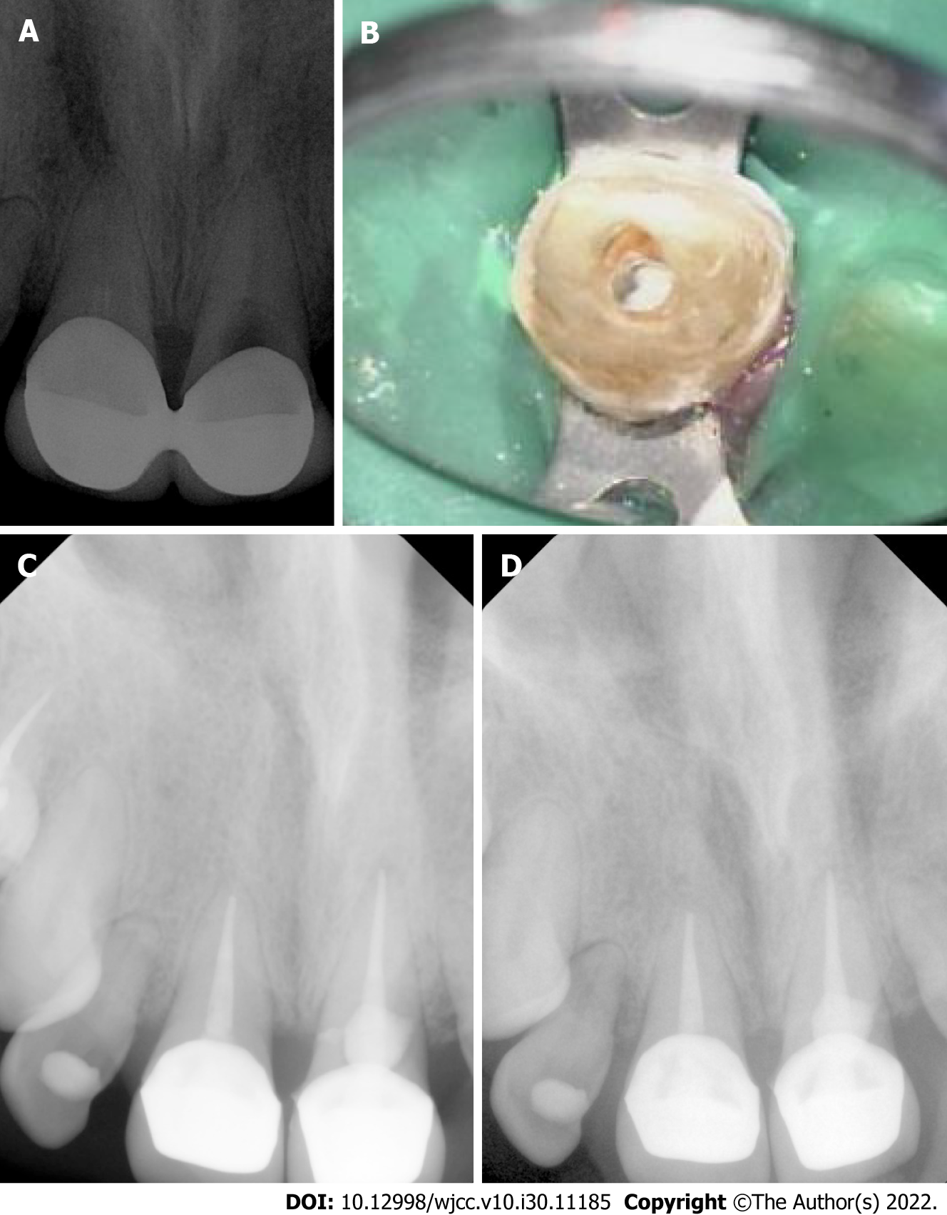
Figure 1 Imaging.
A: Preoperative preapical radiograph showing tooth 21 with large resorption; B: Clinical image showing bioceramic material placed in area of resorption; C: Radiograph was taken after endodontic treatment is completed and final restorations placed; D: Month 18 follow up radiograph showed no abnormalities related to teeth 11 and 21.
- Citation: Riyahi AM. Bioceramics utilization for the repair of internal resorption of the root: A case report. World J Clin Cases 2022; 10(30): 11185-11189
- URL: https://www.wjgnet.com/2307-8960/full/v10/i30/11185.htm
- DOI: https://dx.doi.org/10.12998/wjcc.v10.i30.11185


