Copyright
©The Author(s) 2022.
World J Clin Cases. Oct 26, 2022; 10(30): 11122-11127
Published online Oct 26, 2022. doi: 10.12998/wjcc.v10.i30.11122
Published online Oct 26, 2022. doi: 10.12998/wjcc.v10.i30.11122
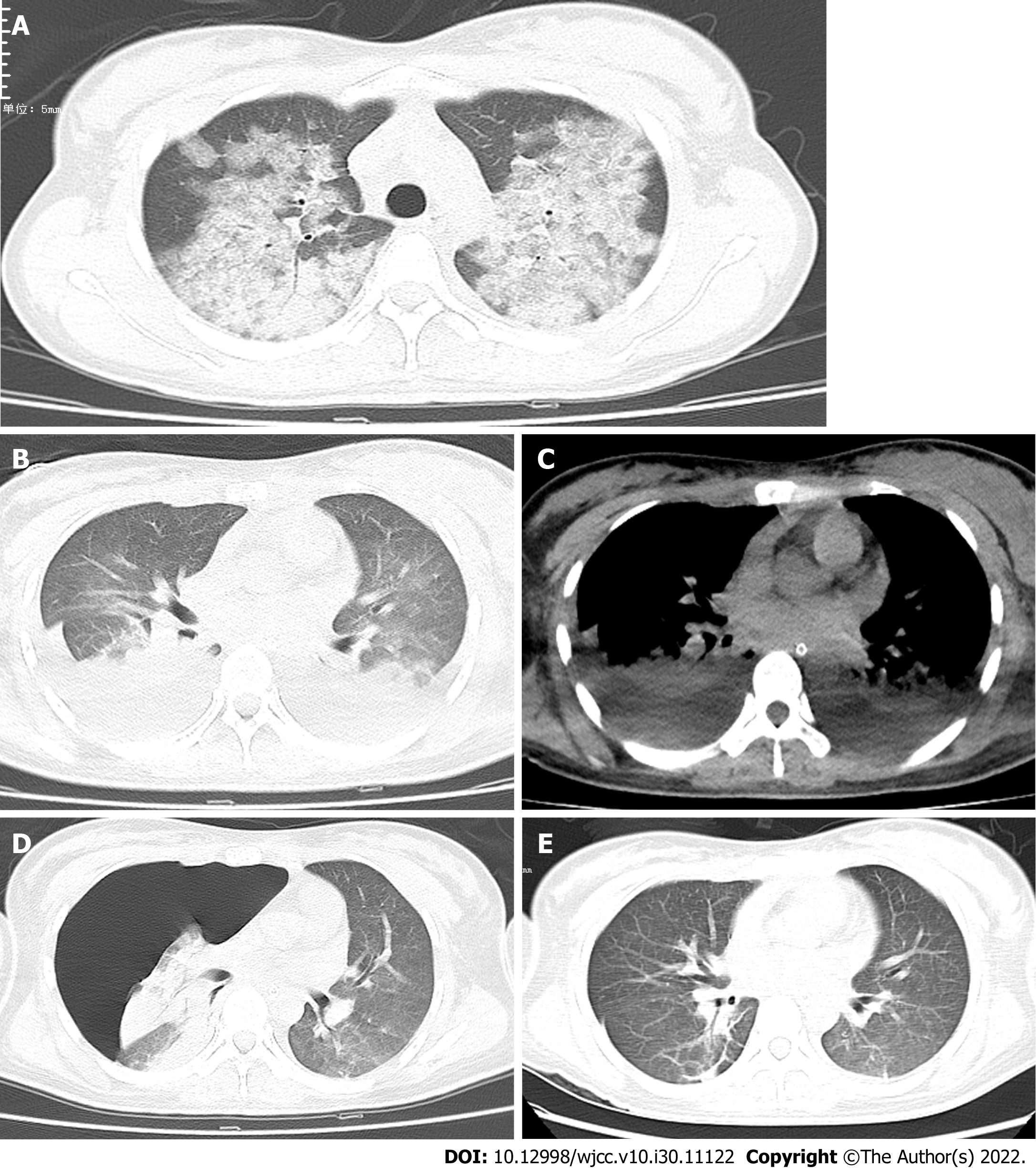
Figure 1 Chest computed tomography scan.
A: On the second day of admission, the computed tomography (CT) scan showed a small pleural effusion and bilateral lung multifocal ground-glass opacity; B and C: On the 7th day, transverse chest CT scan of the lungs revealed large multiple patchy ground-glass opacities with consolidation (B), and a possible large pleural effusion in both lungs (C); D: On the 11th day, transverse chest CT scan showed significant reduction of ground-glass opacities and consolidations complicated with severe right pneumothorax; E: On the 14th day, chest CT scan suggested the disappearance of pneumothorax.
- Citation: Cai ZY, Xu BP, Zhang WH, Peng HW, Xu Q, Yu HB, Chu QG, Zhou SS. Acute respiratory distress syndrome following multiple wasp stings treated with extracorporeal membrane oxygenation: A case report. World J Clin Cases 2022; 10(30): 11122-11127
- URL: https://www.wjgnet.com/2307-8960/full/v10/i30/11122.htm
- DOI: https://dx.doi.org/10.12998/wjcc.v10.i30.11122


