Copyright
©The Author(s) 2022.
World J Clin Cases. Oct 26, 2022; 10(30): 11037-11043
Published online Oct 26, 2022. doi: 10.12998/wjcc.v10.i30.11037
Published online Oct 26, 2022. doi: 10.12998/wjcc.v10.i30.11037
Figure 1 The clinical characteristics of the case.
A: The blue arrow shows the fistula presenting as a neoplasm located in the right hip of the patient. The scar with round features is indicated by the yellow arrow; B: The blue arrow shows the duplicated tubular structure, which is dilated and full of fecal slag-like secretion; C: Abdominal computed tomography scan reveals the double ureter (yellow arrow), compressed uterus (white arrow), duplicated tubular structure (blue arrow), and contrast agent appearing in the fistula (green arrow); D: The three-dimensional reconstruction image shows abnormal morphology of the sacrum (blue arrow) and absence of some bone structures; E: Radiograph of the right foot shows pes cavus.
Figure 2 Images captured during the surgery demonstrating the process of treatment.
A: Removing the fistula in the hip, after methylene blue staining; B: The duplicated tubular structure was removed from the abdominal pelvic cavity; C: The double ureter located during the operation (blue arrow); D: Freeing the duplicated colon. The blue arrow indicates the narrow pelvic floor, while the green arrow shows the complete colonic duplication under laparoscopic view.
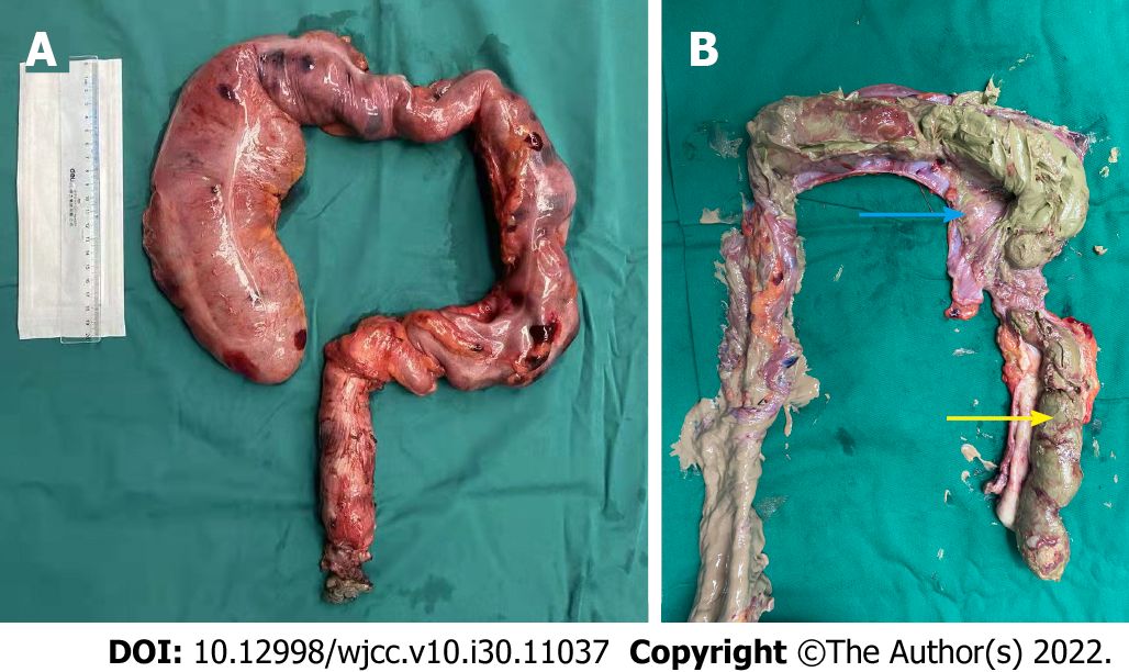
Figure 3 Specimens of the duplicated colon.
A: Complete surgery specimen; B: Mucosa of the duplicated tubular structure indicated by the blue arrow; the content of the lumen is a large amount of fecal slag-like secretion (yellow arrow).
Figure 4 Microscopy of the specimen with hematoxylin and eosin staining.
A: Under 40 × magnification, the layers of the duplicated tubular structure are mucous, muscular mucosae, submucous, muscular, and serosa as shown by black arrows from top to bottom; B: Under 100 × magnification, numerous lymphocytes gather in the mucous layer are presented by the black arrow.
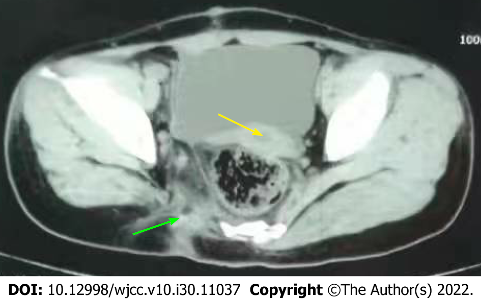
Figure 5 Abdominal pelvic computed tomography review (8 mo postoperatively).
The uterus has almost moved back to its normal position (yellow arrow). The wall of the pelvic floor remains normal in the previous operative region (green arrow), and there is no hernia.
- Citation: Cai X, Bi JT, Zheng ZX, Liu YQ. Complete colonic duplication presenting as hip fistula in an adult with pelvic malformation: A case report. World J Clin Cases 2022; 10(30): 11037-11043
- URL: https://www.wjgnet.com/2307-8960/full/v10/i30/11037.htm
- DOI: https://dx.doi.org/10.12998/wjcc.v10.i30.11037


