Copyright
©The Author(s) 2022.
World J Clin Cases. Oct 16, 2022; 10(29): 10779-10786
Published online Oct 16, 2022. doi: 10.12998/wjcc.v10.i29.10779
Published online Oct 16, 2022. doi: 10.12998/wjcc.v10.i29.10779
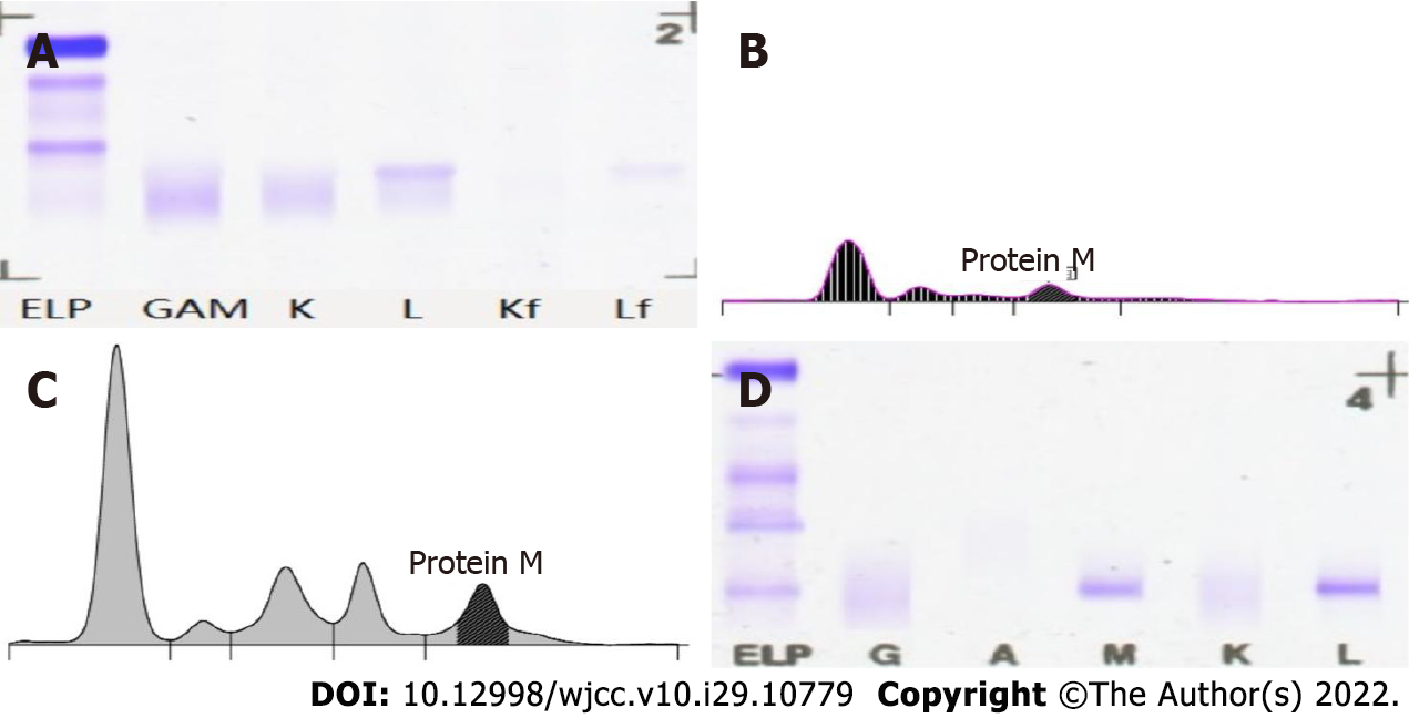
Figure 1 Electrophoretic mapping of the patient on November 30, 2021.
A: Urine Benzedrine electrophoresis; B: Urine protein electrophoresis; C: Serum protein electrophoresis; D: Serum immunofixation electrophoresis. ELP, GAM, K, L, Kf and Lf: Protein electrophoresis/immunofixation lanes.
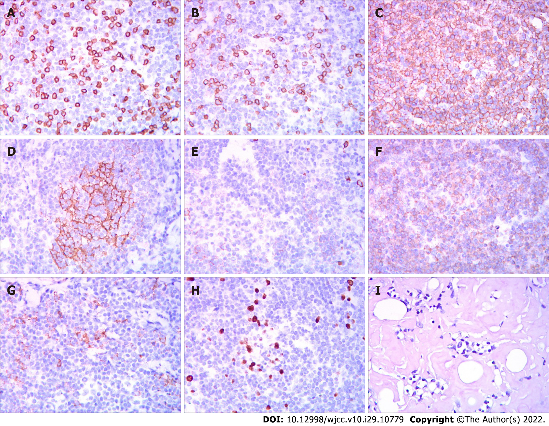
Figure 2 The left cervical lymph node biopsy on December 1, 2021.
A: CD3; B: CD5; C: CD20; D: CD21; E: CD56; F: CD79A; G: CD138; H: Ki-67; I: Congo red.
- Citation: Zhao ZY, Tang N, Fu XJ, Lin LE. Secondary light chain amyloidosis with Waldenström’s macroglobulinemia and intermodal marginal zone lymphoma: A case report. World J Clin Cases 2022; 10(29): 10779-10786
- URL: https://www.wjgnet.com/2307-8960/full/v10/i29/10779.htm
- DOI: https://dx.doi.org/10.12998/wjcc.v10.i29.10779


