Copyright
©The Author(s) 2022.
World J Clin Cases. Oct 16, 2022; 10(29): 10721-10727
Published online Oct 16, 2022. doi: 10.12998/wjcc.v10.i29.10721
Published online Oct 16, 2022. doi: 10.12998/wjcc.v10.i29.10721
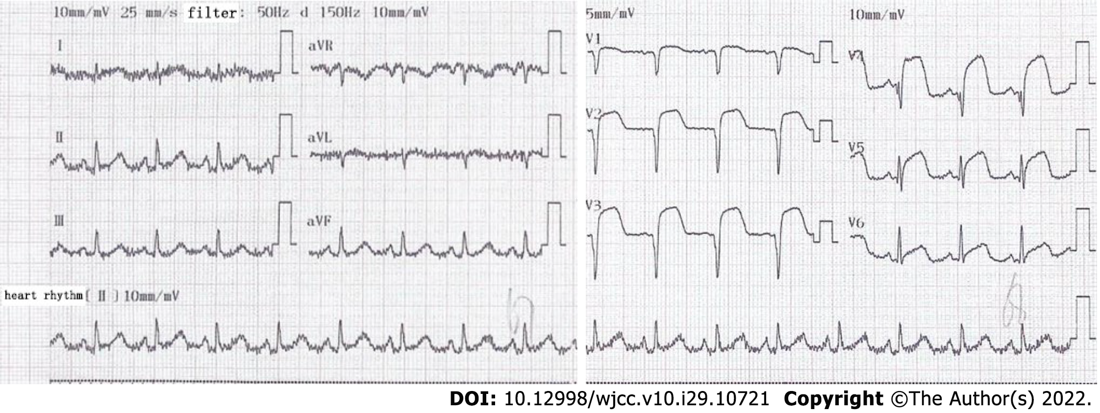
Figure 1 Electrocardiogram showed ST-segment elevation myocardial infarction.
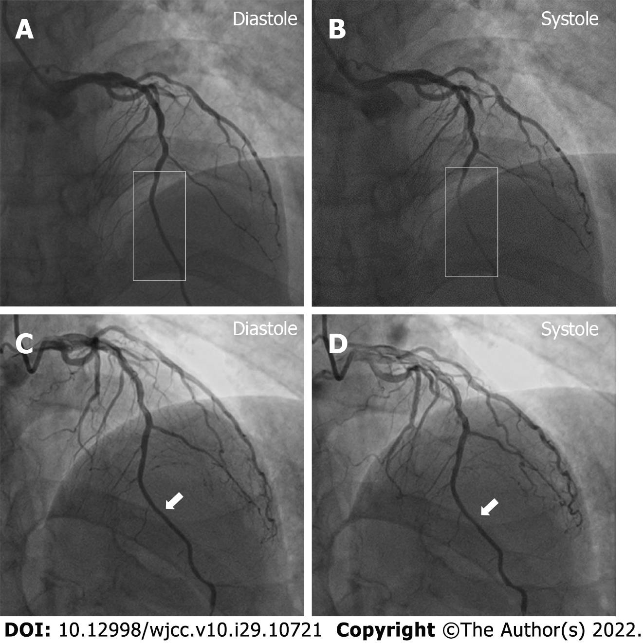
Figure 2 Myocardial bridging was identified on coronary angiography.
A and B: Myocardial bridging (rectangle) was stenosed about 90% during systole and recovered during diastole on January 14, 2020; C and D: The myocardial bridging (white arrow) was stenosed about 30% on October 13, 2020, and its length was reduced during follow-up.
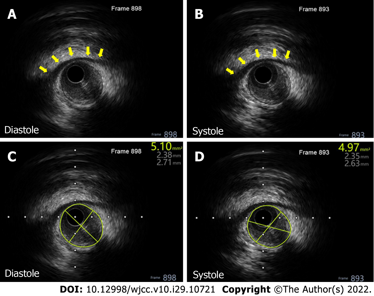
Figure 3 Intravascular ultrasound showed myocardial bridging.
There was no obvious change of the lumen area during systole during follow-up. A and B: An apparent ‘half-moon’ phenomenon of the myocardial bridging on October 13, 2020 (yellow arrow); C and D: The vascular lumen area of the mural coronary artery was 5.10 mm2 during diastole and 4.97 mm2 during systole.
- Citation: Li HH, Liu MW, Zhang YF, Song BC, Zhu ZC, Zhao FH. Myocardial bridging phenomenon is not invariable: A case report. World J Clin Cases 2022; 10(29): 10721-10727
- URL: https://www.wjgnet.com/2307-8960/full/v10/i29/10721.htm
- DOI: https://dx.doi.org/10.12998/wjcc.v10.i29.10721


