Copyright
©The Author(s) 2022.
World J Clin Cases. Oct 16, 2022; 10(29): 10638-10646
Published online Oct 16, 2022. doi: 10.12998/wjcc.v10.i29.10638
Published online Oct 16, 2022. doi: 10.12998/wjcc.v10.i29.10638
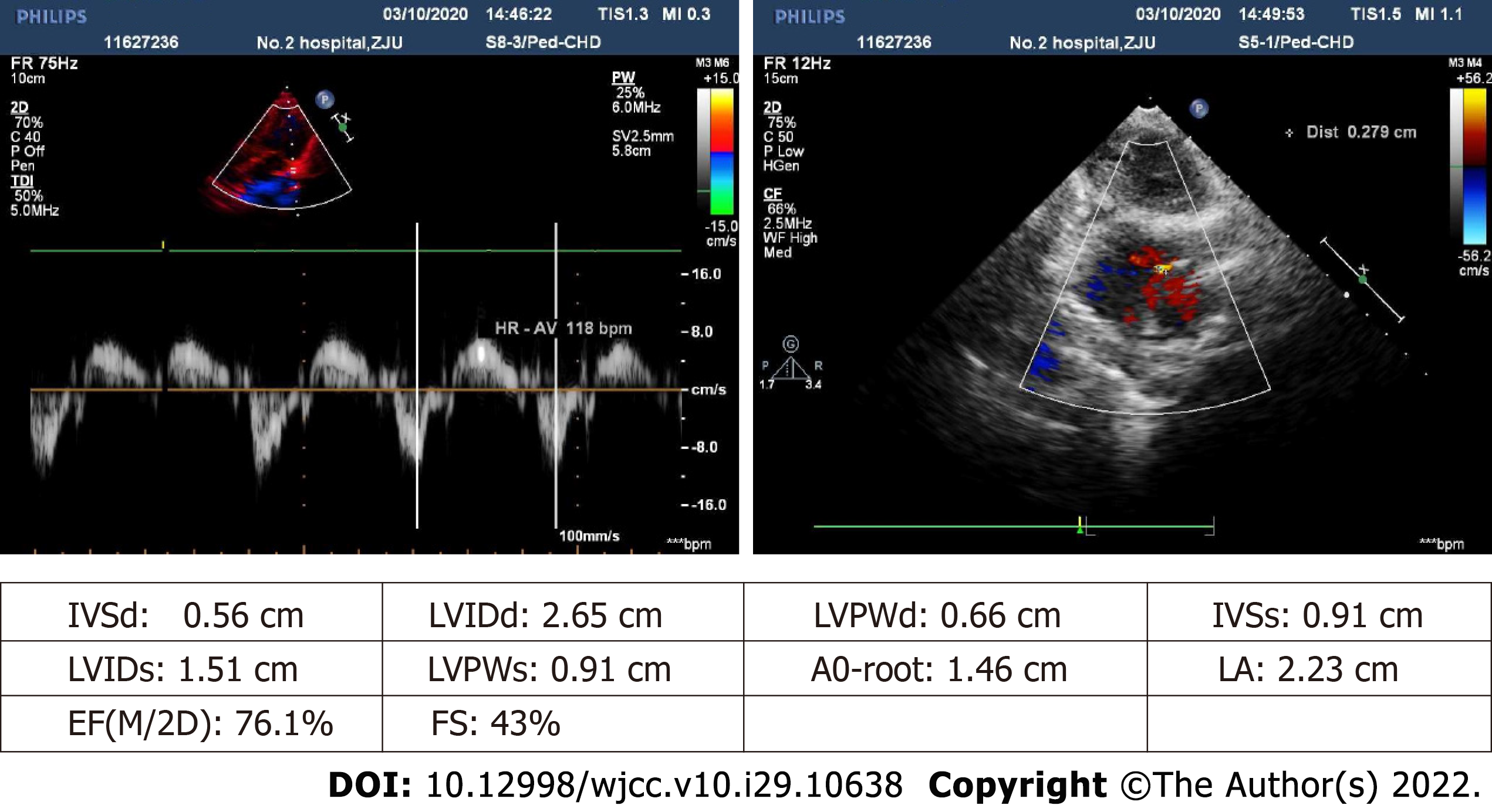
Figure 1 Echocardiographic imaging prior to surgery.
Atrial and ventricular chambers are of normal size (no abnormal internal echoes) and expected thickness, with visible 0.3-cm echoic interruption of middle and lower atrial septum (slightly less than in the prior year). Color Doppler flow imaging (CDFI) reveals left-to-right red-colored streamers, shunt velocity not measured. Interventricular septum remains structurally intact. Valvular echoes appear satisfactory (opening/closing normally), exhibiting trace tricuspid systolic regurgitation (multicolored, mainly blue) by CDFI.
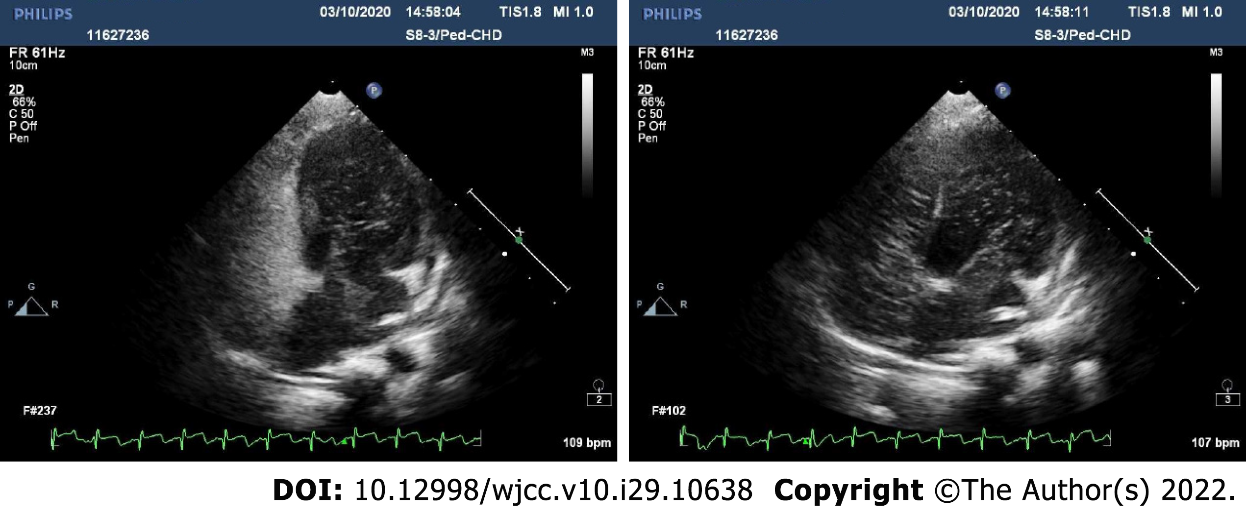
Figure 2 Agitated-saline contrast echocardiography (preoperative images).
Right heart filled well after vibrated normal saline (3 mL) injection of cubital vein. In resting state, few left heart air bubbles formed during third cardiac cycle; whereas many more (> 35 per frame) arose during fifth cardiac cycle, ostensibly from left and right upper pulmonary veins. Valsalva maneuver during third cardiac cycle also triggered a flurry of left heart bubbles (> 35/frame). These findings suggest atrial-level right-left shunting, pulmonary arteriovenous fistula not excluded.
- Citation: Liu L, Chen P, Fang LL, Yu LN. Perioperative anesthesia management in pediatric liver transplant recipient with atrial septal defect: A case report. World J Clin Cases 2022; 10(29): 10638-10646
- URL: https://www.wjgnet.com/2307-8960/full/v10/i29/10638.htm
- DOI: https://dx.doi.org/10.12998/wjcc.v10.i29.10638


