Copyright
©The Author(s) 2022.
World J Clin Cases. Oct 16, 2022; 10(29): 10622-10628
Published online Oct 16, 2022. doi: 10.12998/wjcc.v10.i29.10622
Published online Oct 16, 2022. doi: 10.12998/wjcc.v10.i29.10622
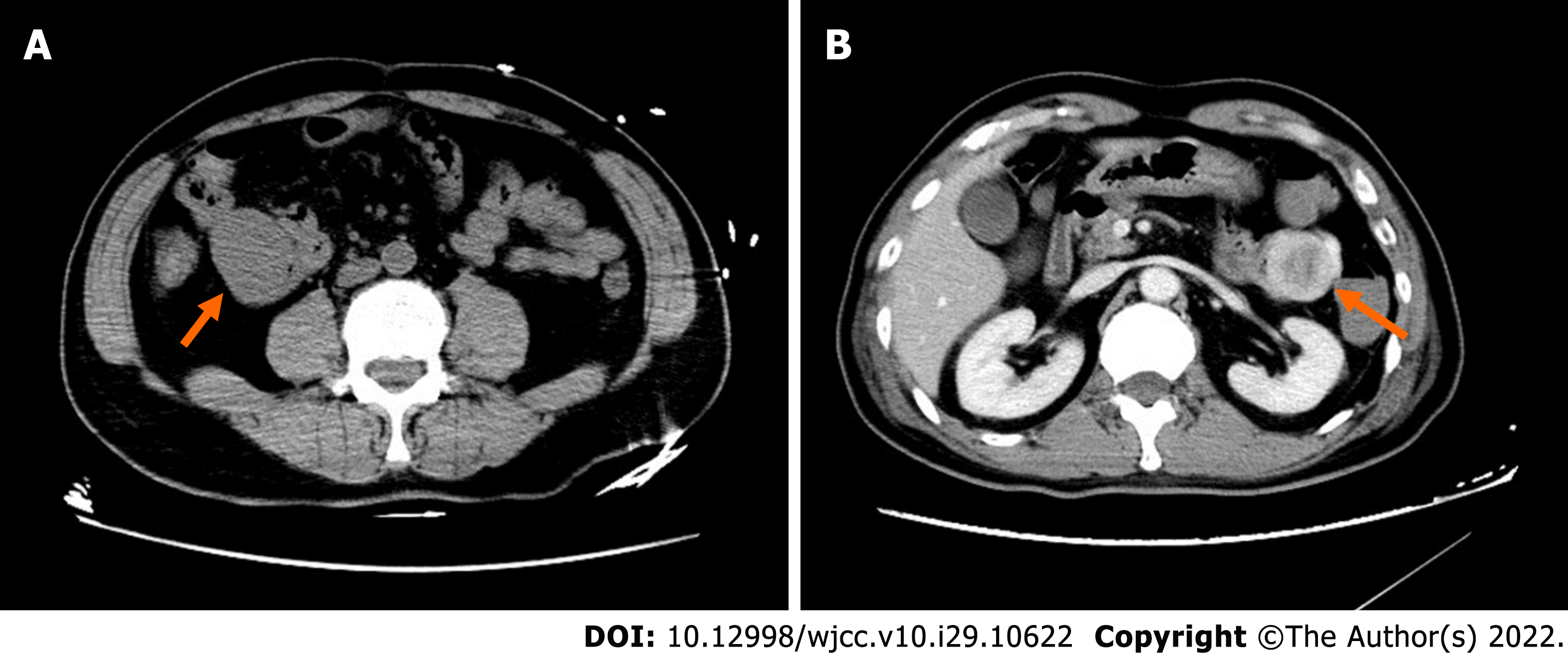
Figure 1 Computed tomography imaging.
A: A 68-year-old man with a wandering small intestinal stromal tumor. At the initial visit, non-contrast computed tomography (CT) scan shows a 4.1 cm × 3.3 cm × 5.0 cm mass located below the right kidney in the right upper abdomen and anterior border of the major lumbar muscle, and the lesion is not clearly demarcated from the small intestine; B: Two days later, contrast-enhanced CT shows the mass (4.6 cm × 3.4 cm × 5.0 cm) in the upper left abdomen in front of the left kidney with non-homogenous enhancement and a thickened artery visible at the edge of the mass. The mass on right upper abdomen cannot be visualized.
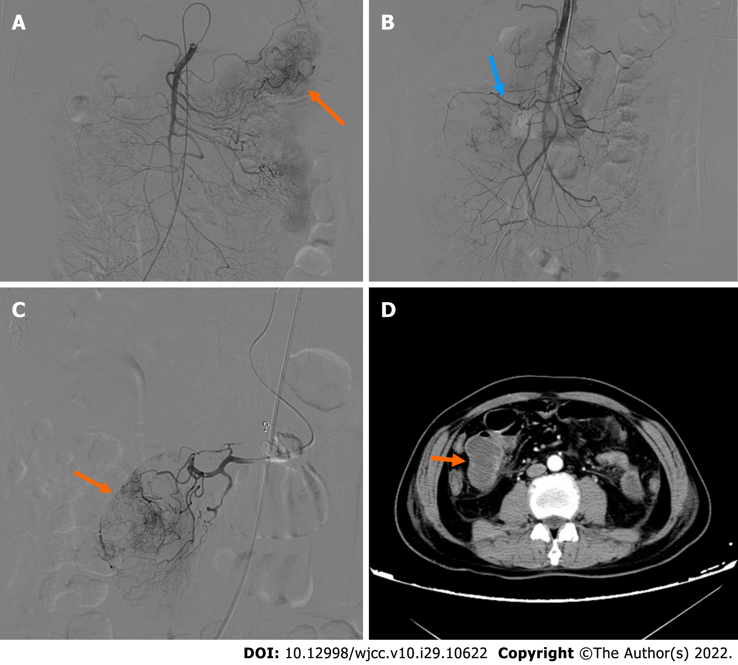
Figure 2 Digital subtraction angiography imaging.
A: First digital subtraction angiography (DSA) showing the mass located in the left abdomen. The distal vascular branches of the first branch of the jejunum appear to be abundant and haphazard with contrast media exudation and retention in the intestinal lumen; B: Second DSA shows the first and second branches of the jejunum in the right abdomen; C: Superselective arteriography showing tumor staining; D: Contrast-enhanced computed tomography after the second embolization showing the mass in the right mid-abdomen.
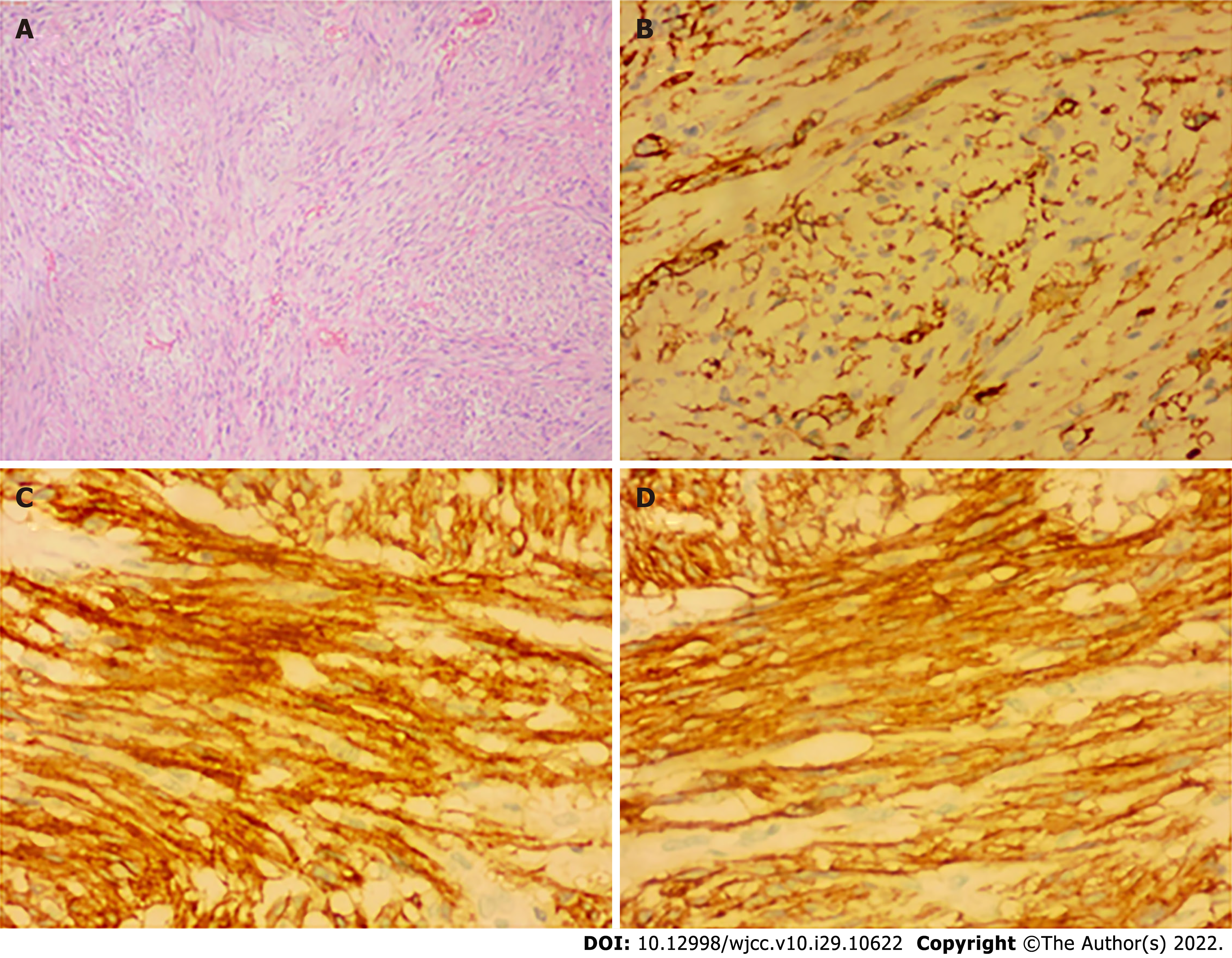
Figure 3 Histopathological and immunohistochemical.
A: Histopathological examination of the resected specimen (hematoxylin and eosin; × 40) showing submucosal stromal tumor with intact capsule, infarction, spindle shaped cells, and nuclear division less than 1/10 hibernation promoting factor. Immunohistochemical test results revealed the following (× 200); B: CD34+; C: CD117+; D: Dog1+.
- Citation: Su JZ, Fan SF, Song X, Cao LJ, Su DY. Wandering small intestinal stromal tumor: A case report. World J Clin Cases 2022; 10(29): 10622-10628
- URL: https://www.wjgnet.com/2307-8960/full/v10/i29/10622.htm
- DOI: https://dx.doi.org/10.12998/wjcc.v10.i29.10622


