Copyright
©The Author(s) 2022.
World J Clin Cases. Oct 16, 2022; 10(29): 10535-10542
Published online Oct 16, 2022. doi: 10.12998/wjcc.v10.i29.10535
Published online Oct 16, 2022. doi: 10.12998/wjcc.v10.i29.10535
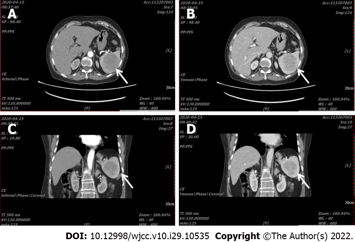
Figure 1 Abdominal computed tomography angiograph scan of Case 2.
A: (arterial phase) and B (venous phase): In transverse section, A pre-operative computed tomography (CT) scan revealed splenomegaly, and a tumor (6.0 cm × 5.7 cm in size) with a CT value 48 HU showing gradual enhancement; C (arterial phase) and D (venous phase): The splenic tumor is shown in the coronal phase. White arrow indicates the tumor.
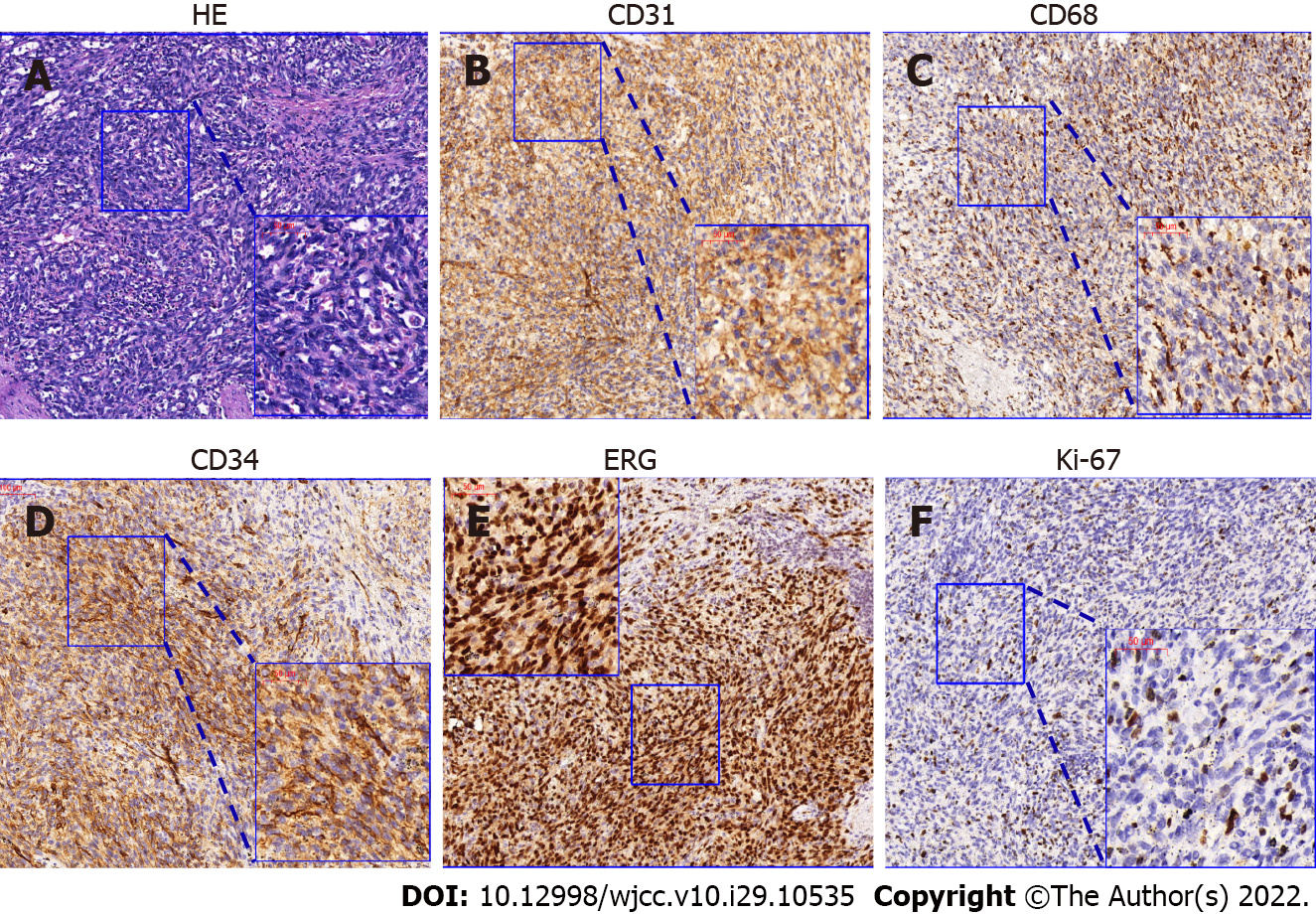
Figure 2 HE and immunohistochemical characteristics of Case 1.
A: The littoral cell angiosarcoma contains perivascular sinus-like heterocysts with dark nuclei and multiple mitotic phases on HE staining; B and C: Immunohistochemical phenotype analysis showed that the tumor cells were CD31 positive, while CD68 was focally positive; D and E: Furthermore, typical endothelial markers CD34 and ERG were positive and expressed in perivascular cells in littoral cell angiosarcoma; F: The Ki-67 index was 5%-10% (black arrow).
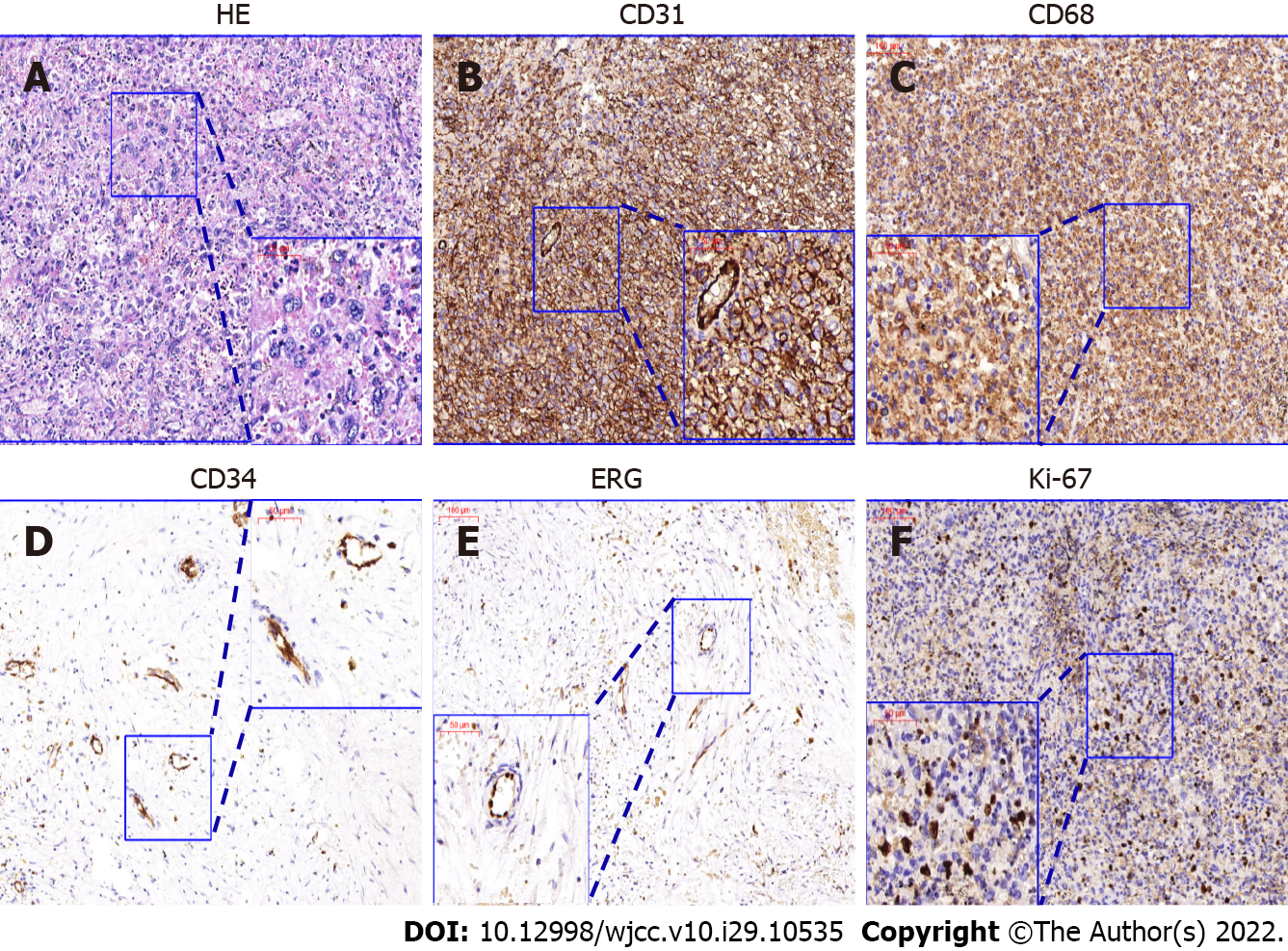
Figure 3 HE and immunohistochemical characteristics of Case 2.
A: The tumor in Case 2 contained numerous large cells with abundant blue cytoplasm with binucleated and trinucleated cells, which is coincident with the characteristics of histiocytic sarcoma; B and C: Immunohistochemical phenotype analysis showed that tumor cells were CD31 positive, while CD68 was generally positive; D and E: CD34 and ERG were only positively expressed in vascular endothelial cells; F: The Ki-67 index was 15%-20%.
- Citation: Luo H, Wang T, Xiao L, Wang C, Yi H. Multiple disciplinary team management of rare primary splenic malignancy: Two case reports. World J Clin Cases 2022; 10(29): 10535-10542
- URL: https://www.wjgnet.com/2307-8960/full/v10/i29/10535.htm
- DOI: https://dx.doi.org/10.12998/wjcc.v10.i29.10535


