Copyright
©The Author(s) 2022.
World J Clin Cases. Sep 6, 2022; 10(25): 8962-8967
Published online Sep 6, 2022. doi: 10.12998/wjcc.v10.i25.8962
Published online Sep 6, 2022. doi: 10.12998/wjcc.v10.i25.8962
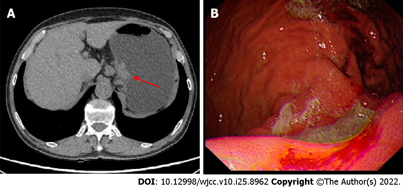
Figure 1 Computer tomography image and gastroscopic picture of initial diagnosis.
A: Computer tomography scan showed local thickening of the gastric body wall; B: Endoscopic image revealed a 3 cm × 3 cm ulcer with irregular borders, mucosal sclerosis and hemorrhagic tendency was revealed on the lesser curvature of the gastric body by gastroscopy.
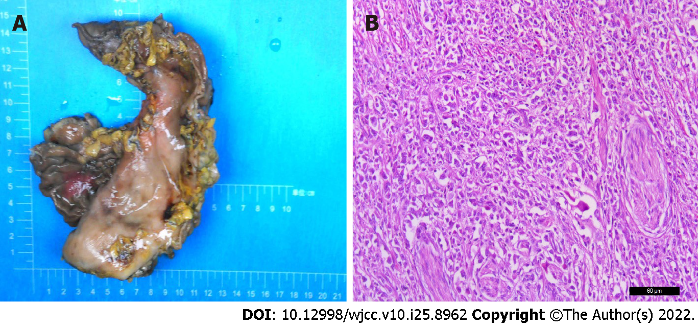
Figure 2 Gross specimen and pathological picture.
A: Inspection of the gastrectomy specimen revealed a tumor measuring 4 cm × 4 cm × 1 cm; B: Hematoxylin-eosin staining of biopsy specimens indicated lymphoepithelioma-like gastric carcinoma (× 200 magnification).
Figure 3 Computer tomography images before and after treatment.
A: Computer tomography before immunotherapy showed metastases to retroperitoneal lymph node and adrenal gland; B: Re-examination after immunotherapy revealed a significant reduction in the size of the metastases.
- Citation: Cui YJ, Ren YY, Zhang HZ. Treatment of gastric carcinoma with lymphoid stroma by immunotherapy: A case report. World J Clin Cases 2022; 10(25): 8962-8967
- URL: https://www.wjgnet.com/2307-8960/full/v10/i25/8962.htm
- DOI: https://dx.doi.org/10.12998/wjcc.v10.i25.8962


