Copyright
©The Author(s) 2022.
World J Clin Cases. Jul 6, 2022; 10(19): 6744-6749
Published online Jul 6, 2022. doi: 10.12998/wjcc.v10.i19.6744
Published online Jul 6, 2022. doi: 10.12998/wjcc.v10.i19.6744
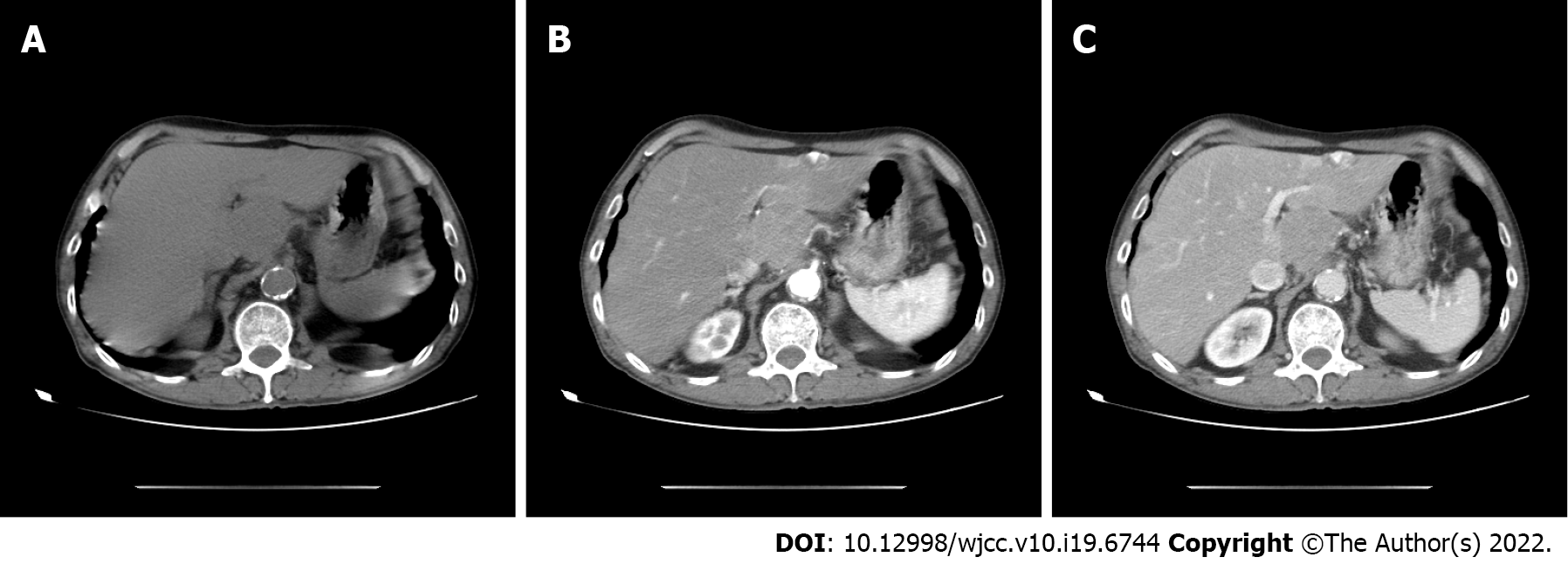
Figure 1 Abdominal computed tomography shows a 3.
0 cm x 3.5 cm irregular tumor, with uneven density, and mild enhancement in the arterial phase in the left caudate lobe. A: Preoperative computed tomography (CT) plain scan period; B: Preoperative CT arterial phase; C: Preoperative CT venous phase.
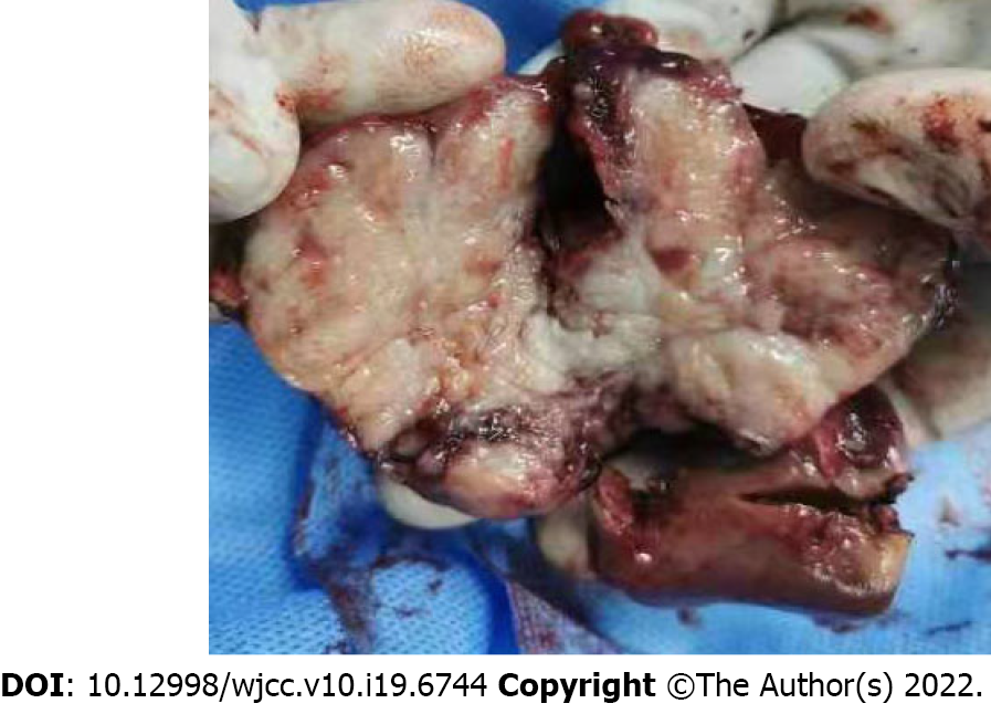
Figure 2 Gross features of the hepatic tumor.
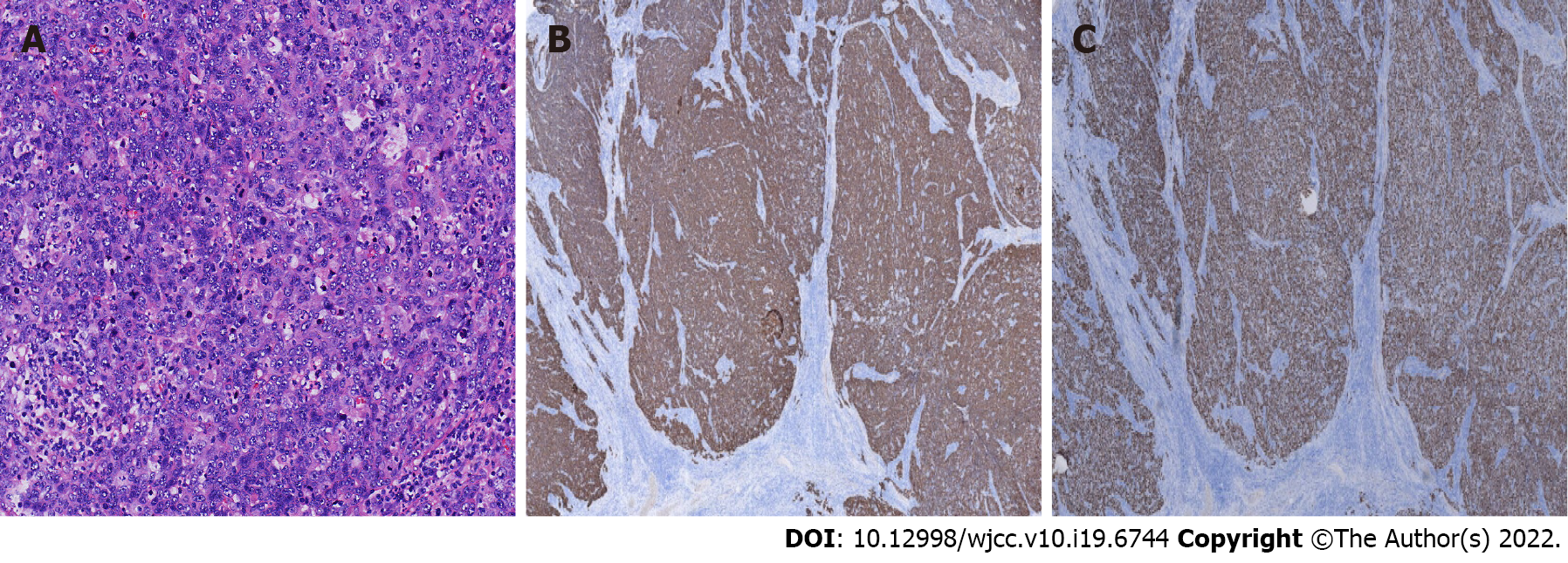
Figure 3 Pathologic characteristicsof the resected liver tumor.
A: The tumor was composed of non-keratinized squamous cells with some keratinization (HE x 200); B: Immunohistochemical findings (IHC x 200); C: Squamous cells expressing strong positive P40 staining (IHC x 200).
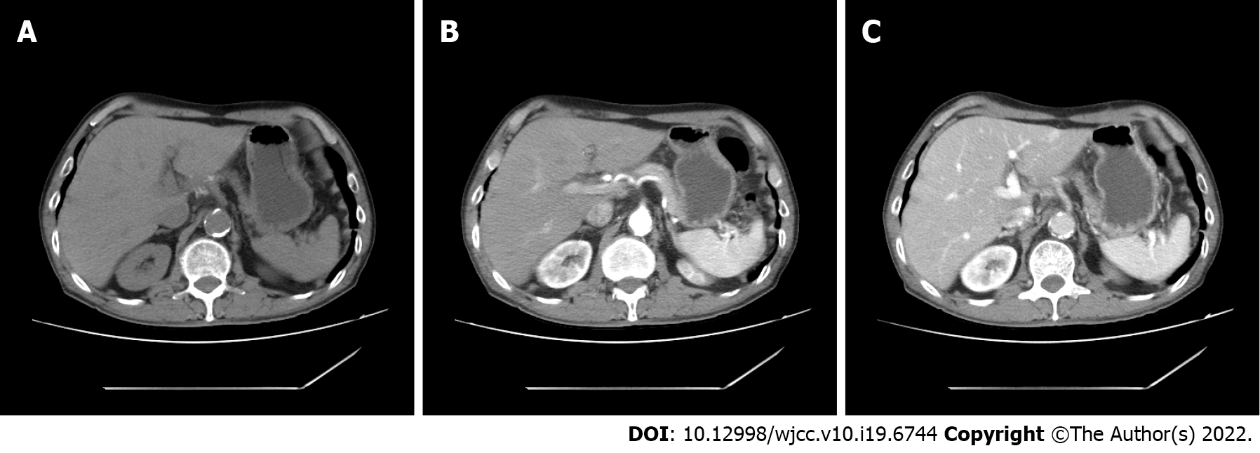
Figure 4 Computed tomography showed no tumor recurrence or metastasis at the resection site.
A: Postoperative computed tomography (CT) plain scan period; B: Postoperative CT arterial phase; C: Postoperative CT venous phase.
- Citation: Kang LM, Yu DP, Zheng Y, Zhou YH. Primary squamous cell carcinoma of the liver: A case report. World J Clin Cases 2022; 10(19): 6744-6749
- URL: https://www.wjgnet.com/2307-8960/full/v10/i19/6744.htm
- DOI: https://dx.doi.org/10.12998/wjcc.v10.i19.6744


