Copyright
©The Author(s) 2022.
World J Clin Cases. Jul 6, 2022; 10(19): 6656-6663
Published online Jul 6, 2022. doi: 10.12998/wjcc.v10.i19.6656
Published online Jul 6, 2022. doi: 10.12998/wjcc.v10.i19.6656
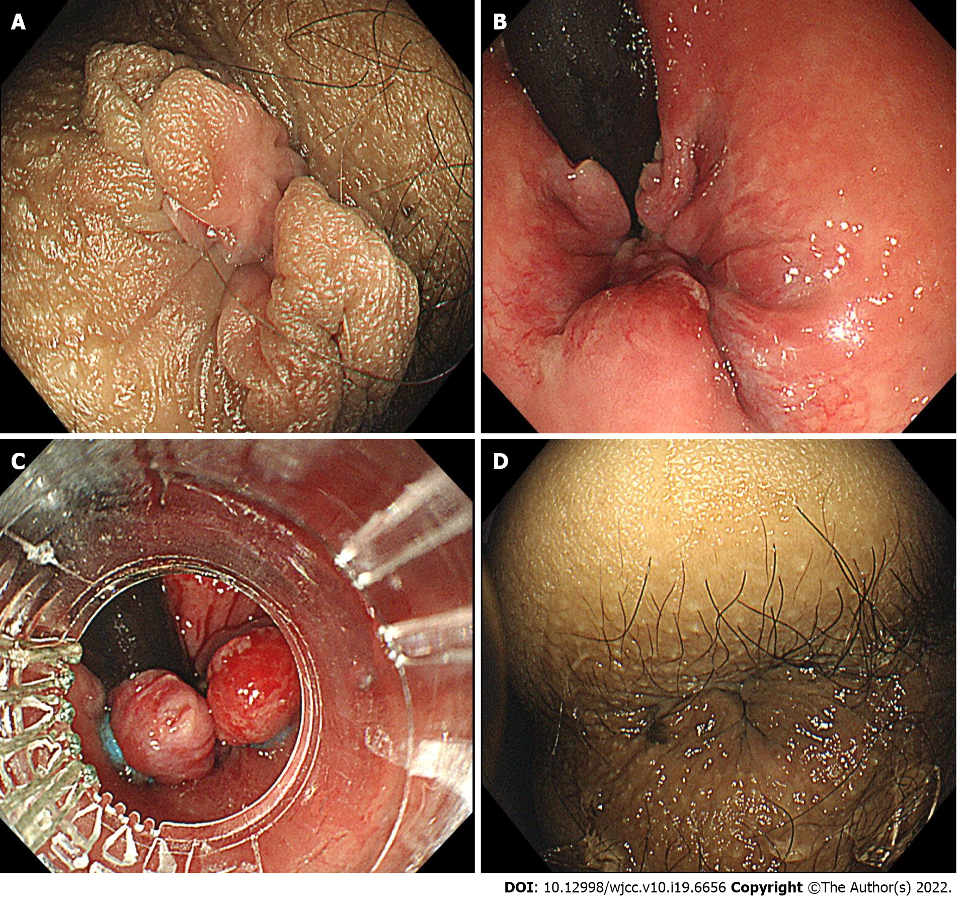
Figure 1 Endoscopic images of endoscopic rubber band ligation therapy.
A: Prolapsed hemorrhoids in forward view before endoscopic rubber band ligation (ERBL); B: Retroflexion view of hemorrhoids before ERBL; C: Ligation of hemorrhoids during ERBL; D: the prolapsed hemorrhoid tissue was withdrawn into the anus after ERBL.
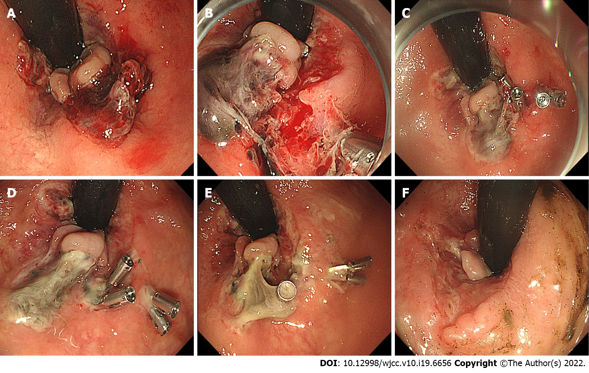
Figure 2 Endoscopic images during follow-up study.
A: Active oozing of blood on 7 d post-endoscopic rubber band ligation (ERBL); B: Ulcer and active bleeding on 9 d post-ERBL; C: image after endoscopic hemostasis using clips; D: 12 d post-ERBL; E: 15 d post-ERBL; F: 3 mo post-ERBL.
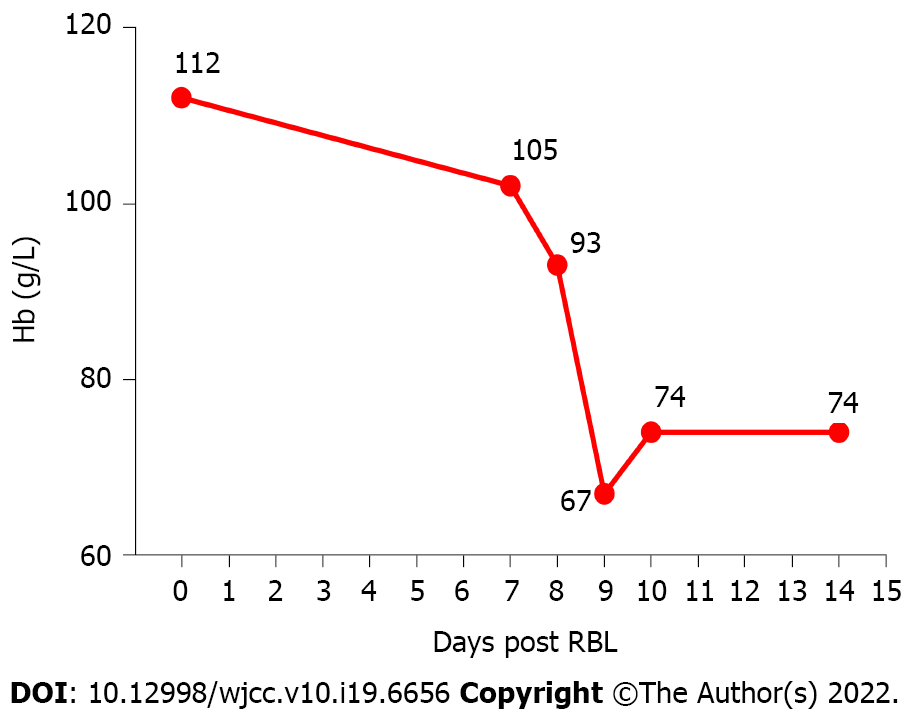
Figure 3 Curve of hemoglobin before and after endoscopic rubber band ligation therapy.
- Citation: Jiang YD, Liu Y, Wu JD, Li GP, Liu J, Hou XH, Song J. Massive gastrointestinal bleeding after endoscopic rubber band ligation of internal hemorrhoids: A case report. World J Clin Cases 2022; 10(19): 6656-6663
- URL: https://www.wjgnet.com/2307-8960/full/v10/i19/6656.htm
- DOI: https://dx.doi.org/10.12998/wjcc.v10.i19.6656


