Copyright
©The Author(s) 2022.
World J Clin Cases. Jul 6, 2022; 10(19): 6647-6655
Published online Jul 6, 2022. doi: 10.12998/wjcc.v10.i19.6647
Published online Jul 6, 2022. doi: 10.12998/wjcc.v10.i19.6647
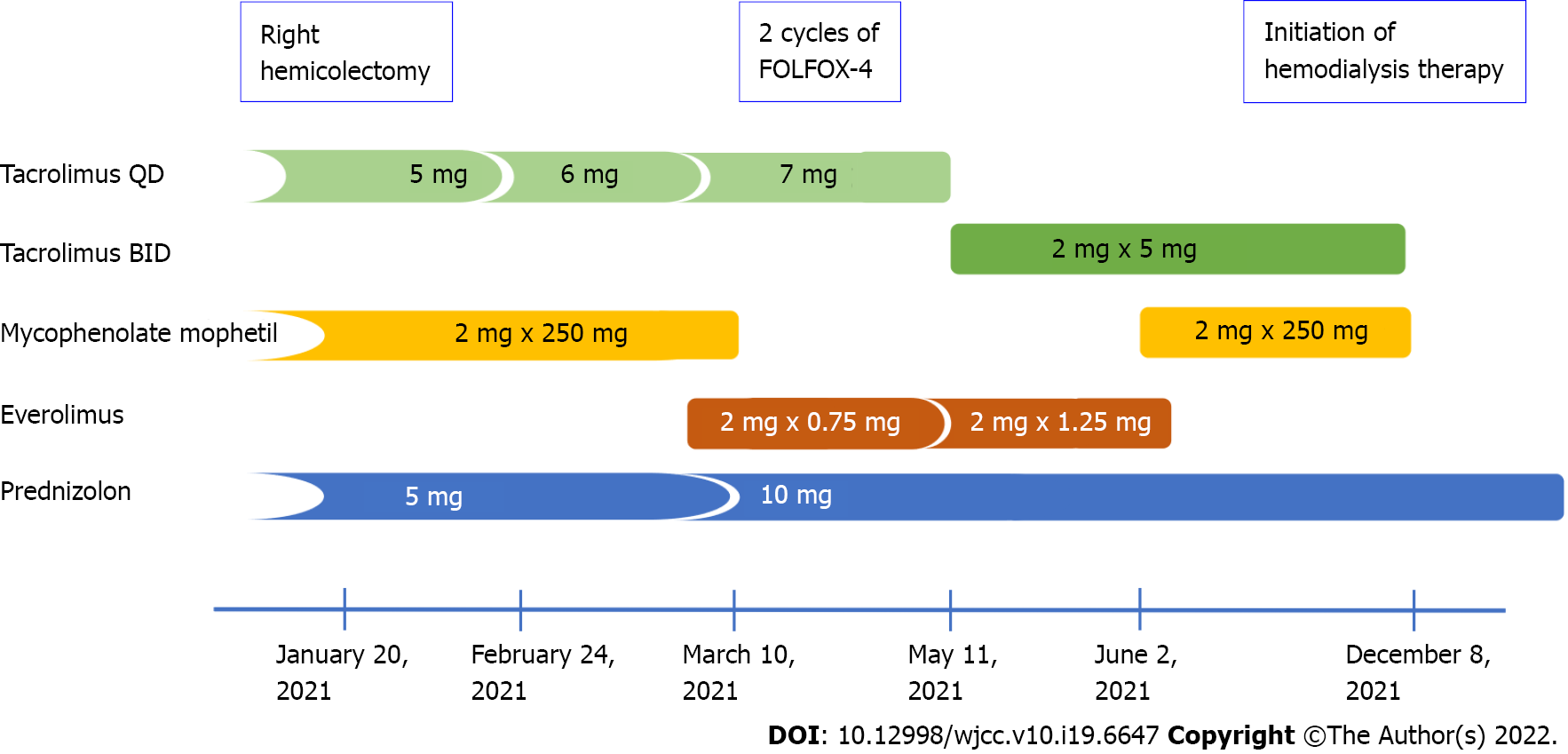
Figure 1 Changes in the immunosuppression therapy after hemicolectomy and diagnosis of colon cancer.
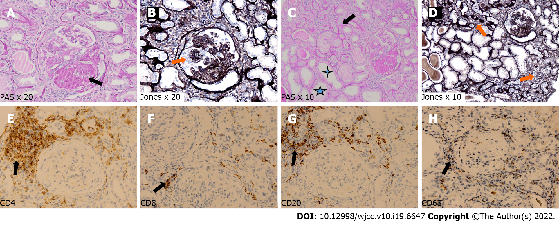
Figure 2 Pathological findings in transplanted kidney biopsy.
A and B: Glomerulus with segmental sclerosis – black arrow (A) – and reactive proliferation of podocytes around sclerosed segment – orange arrow (B); C and D: Area of tubular atrophy – black arrow – and compensative tubular hypertrophy – asterisks (C), with areas of interstitial fibrosis with infiltration of mononuclear cells – orange arrows (D); E–H: Immunohistochemistry showing focal interstitial infiltrates mainly composed of CD4-positive (E) and CD20-positive (G) cells, and diffuse interstitial infiltrates of CD8-positive (F) and CD68-positive (H) cells.
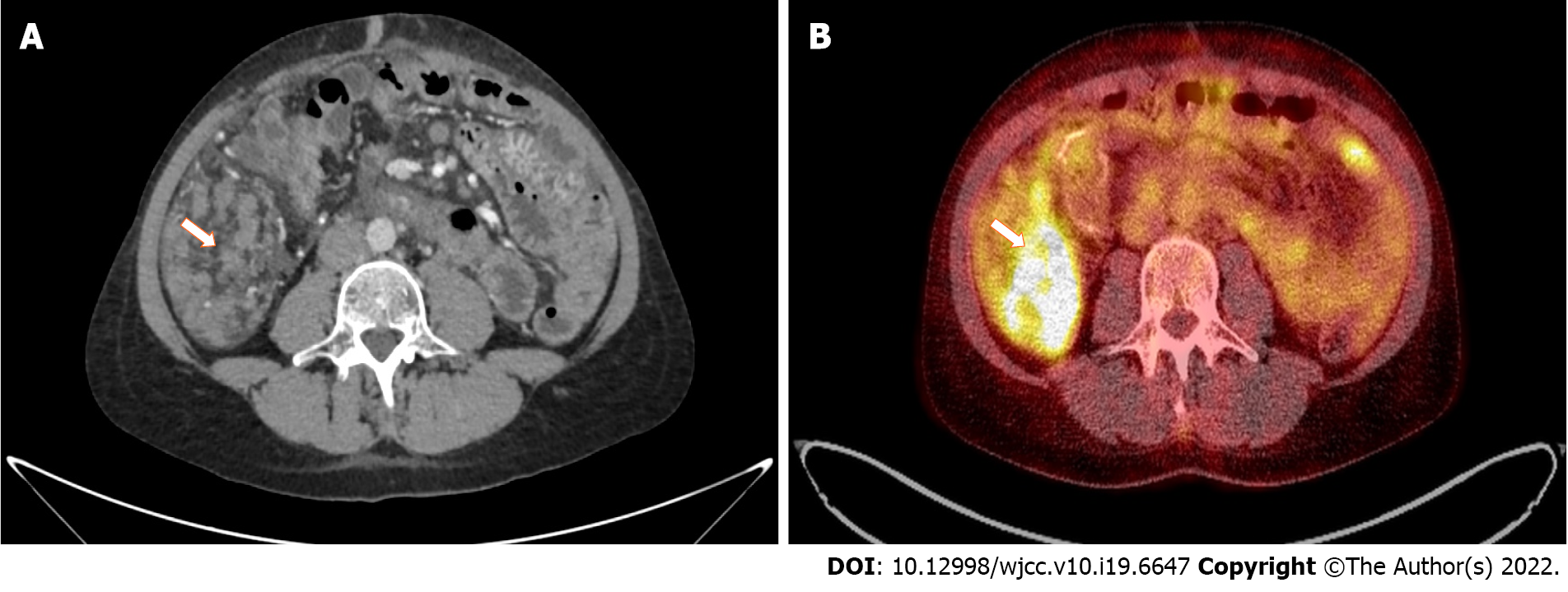
Figure 3 Pathological intraperitoneal infiltration (arrows) measuring 83 mm × 46 mm × 52 mm, with a standardized maximum uptake of 8.
5 located in the right epigastric region, shown in computed tomography (A) and positron emission tomography (B) examinations.
- Citation: Pośpiech M, Kolonko A, Nieszporek T, Kozak S, Kozaczka A, Karkoszka H, Winder M, Chudek J. Transplanted kidney loss during colorectal cancer chemotherapy: A case report. World J Clin Cases 2022; 10(19): 6647-6655
- URL: https://www.wjgnet.com/2307-8960/full/v10/i19/6647.htm
- DOI: https://dx.doi.org/10.12998/wjcc.v10.i19.6647


