Copyright
©The Author(s) 2022.
World J Clin Cases. Jul 6, 2022; 10(19): 6636-6646
Published online Jul 6, 2022. doi: 10.12998/wjcc.v10.i19.6636
Published online Jul 6, 2022. doi: 10.12998/wjcc.v10.i19.6636
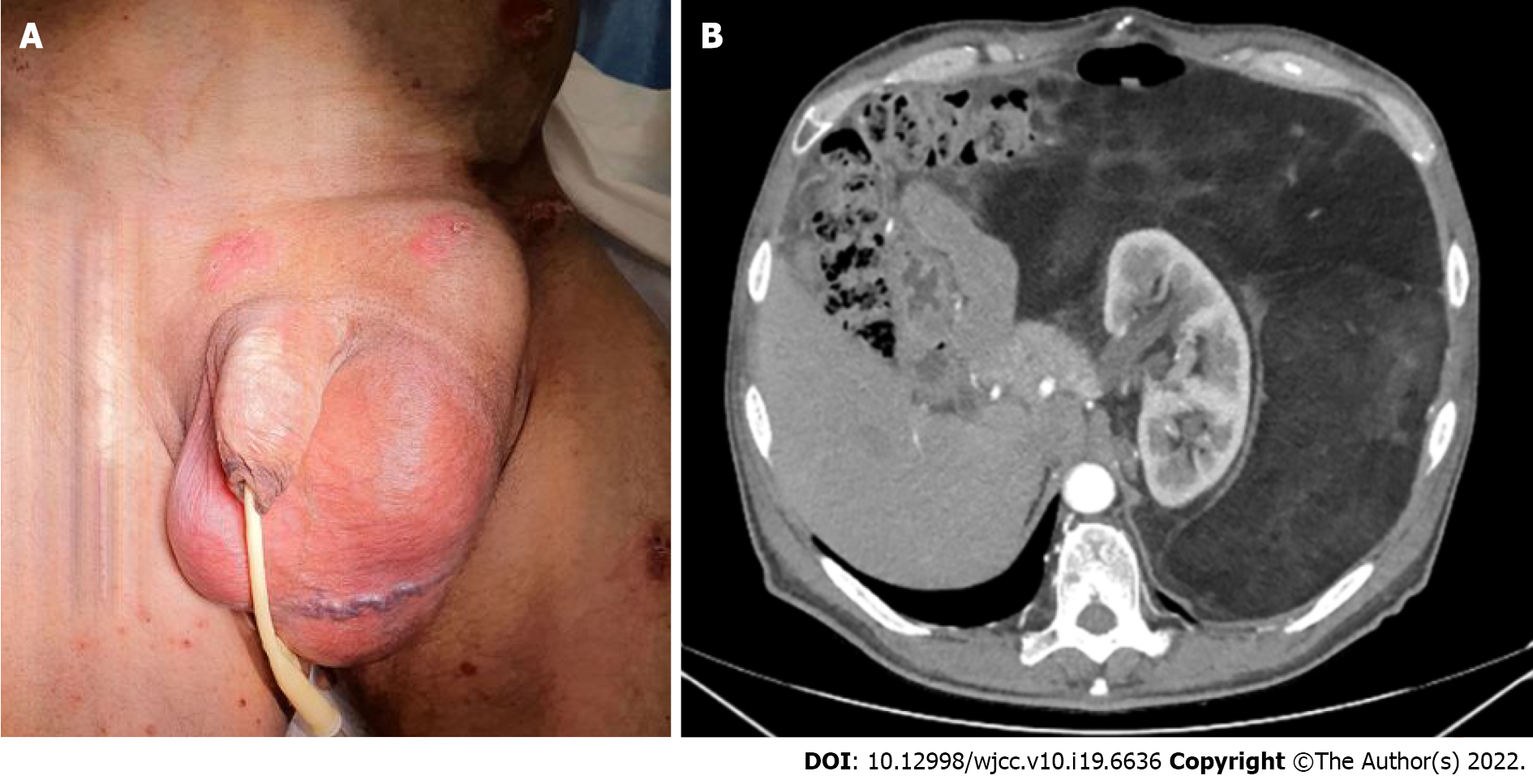
Figure 1 Preoperative image and computed tomography scan.
A: Preoperative image, the patient was initially diagnosed with a left inguinal hernia; B: Preoperative computed tomography scan. A giant retroperitoneal liposarcoma occupied the entire left abdominal cavity with extreme lateralization of the bowel and the left kidney. A grade III hydronephrosis was evident.
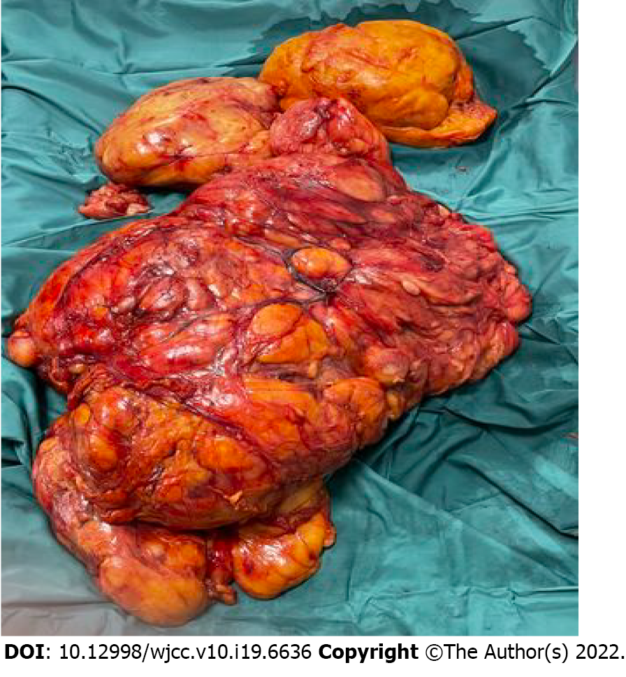
Figure 2 Surgical specimen.
A completely encapsulated liposarcoma was entirely removed without any surrounding organ damage.
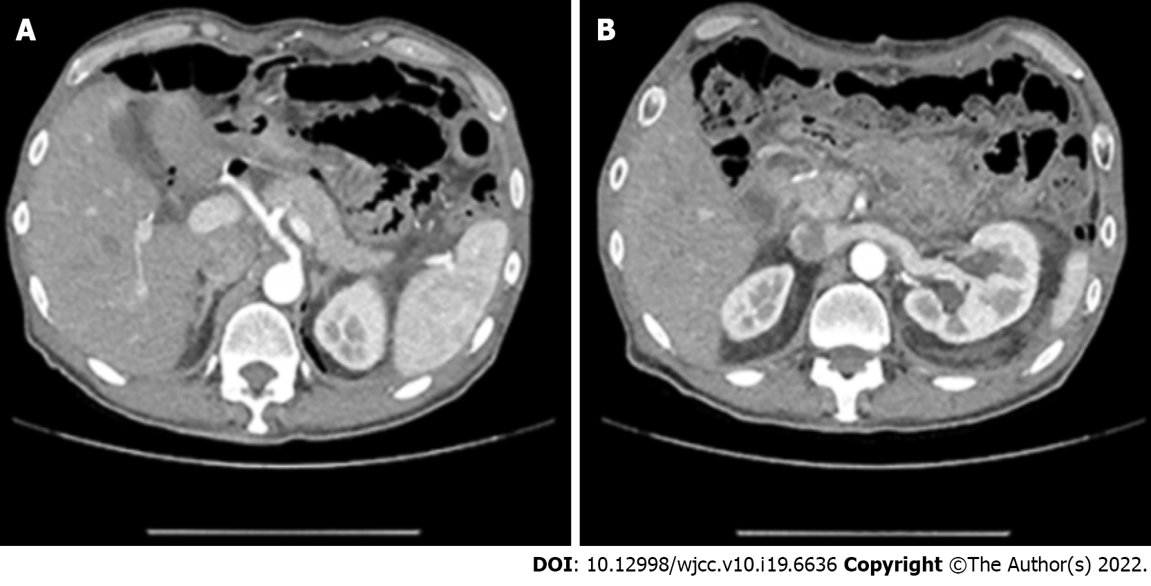
Figure 3 Postoperative computed tomography scan.
The intra-abdominal organs resumed their physiological positions. A: Computed tomography (CT) scan at the level of the celiac trunk; B: CT scan at the level of the left renal vein.
Figure 4 Pathologic picture of well-differentiated liposarcoma.
A: Atypical stromal cells (black arrows; mandatory for diagnosis of well-differentiated liposarcoma) and collagen ropey bands (blue arrow; enhance diagnostic confidence) are present; B: Lipoblastic cell (green arrow).
- Citation: Lieto E, Cardella F, Erario S, Del Sorbo G, Reginelli A, Galizia G, Urraro F, Panarese I, Auricchio A. Giant retroperitoneal liposarcoma treated with radical conservative surgery: A case report and review of literature. World J Clin Cases 2022; 10(19): 6636-6646
- URL: https://www.wjgnet.com/2307-8960/full/v10/i19/6636.htm
- DOI: https://dx.doi.org/10.12998/wjcc.v10.i19.6636


