Copyright
©The Author(s) 2022.
World J Clin Cases. Jun 26, 2022; 10(18): 6136-6140
Published online Jun 26, 2022. doi: 10.12998/wjcc.v10.i18.6136
Published online Jun 26, 2022. doi: 10.12998/wjcc.v10.i18.6136
Figure 1 A 56-year-old man with right-sided sigmoid colon carcinoma.
A: Axial post-contrast computed tomography (CT) scan in the arterial phase demonstrates that the stomach (s) and splenic flexure of colon are normal (*); B: Axial post-contrast CT scan in the arterial phase shows the descending colon entering the peritoneum. The sigmoid colon with the tumor (*) is located on the right side of the ascending colon (a); C: Axial post-contrast CT scan in the arterial phase shows the descending colon (d) crossing to the right at the level of the L4 vertebra and continuing as the sigmoid colon (s) on the right. The tumor (*) is located in a redundant right-sided sigmoid colon. The cecum (c) is displaced toward the left at the level of the L4 transverse process instead of the right pelvic region. The inferior mesenteric artery (arrows) is shown running to the right instead of its normal left-sided course.
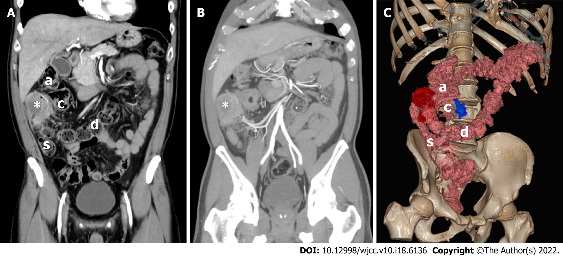
Figure 2 A 56-year-old man with right-sided sigmoid colon carcinoma.
A: Coronal post-contrast computed tomography (CT) scan in the arterial phase demonstrates that the descending colon (d) crosses to the right and continues as the sigmoid colon (s). The tumor (*) is located in a redundant right-sided sigmoid colon occupying the subhepatic region. The ascending colon (a) and the cecum (c) have been displaced; B: Maximum Intensity Projection (MIP) demonstrates that the tumor (*) is located in the right abdomen. The inferior mesenteric artery (arrows) is shown running to the right instead of its normal left-sided course; C: Volume Rendering Technique (VRT) demonstrates abnormal positions of both the sigmoid colon and descending colon. The descending colon (d) is shown crossing to the right and continuing as the sigmoid colon (s). The tumor (red part) is located in a redundant right-sided sigmoid colon. The ascending colon (a) and cecum (c) have been displaced. The ileocecal junction (blue part) is at the L4 level, indicating that the cecum was undescended during embryogenesis.
- Citation: Lyu LJ, Yao WW. Carcinoma located in a right-sided sigmoid colon: A case report. World J Clin Cases 2022; 10(18): 6136-6140
- URL: https://www.wjgnet.com/2307-8960/full/v10/i18/6136.htm
- DOI: https://dx.doi.org/10.12998/wjcc.v10.i18.6136


