Copyright
©The Author(s) 2022.
World J Clin Cases. Jun 26, 2022; 10(18): 6128-6135
Published online Jun 26, 2022. doi: 10.12998/wjcc.v10.i18.6128
Published online Jun 26, 2022. doi: 10.12998/wjcc.v10.i18.6128
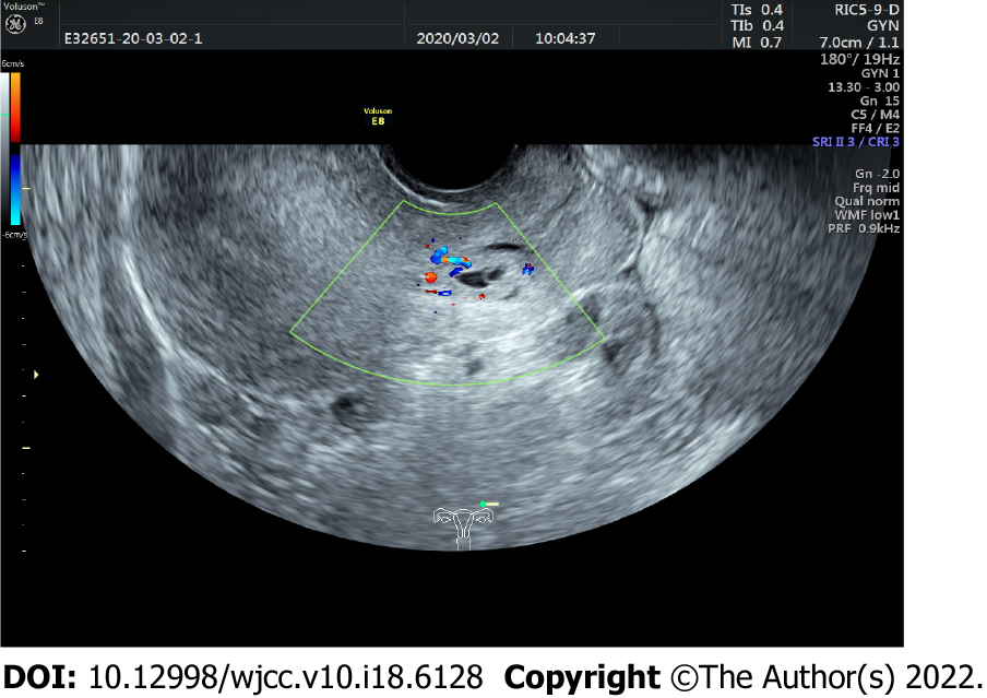
Figure 1 Transvaginal ultrasound image before admission.
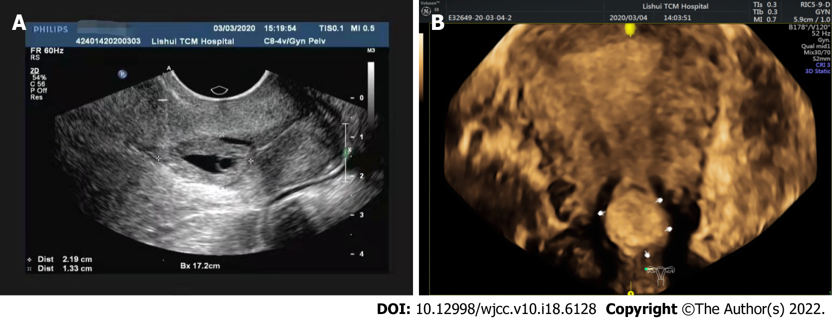
Figure 2 Ultrasound-guided puncture images (A) and after puncture (B).
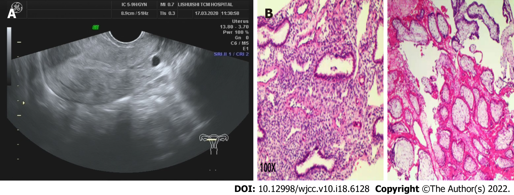
Figure 3 Postoperative ultrasound (A) and pathologic examination (B).
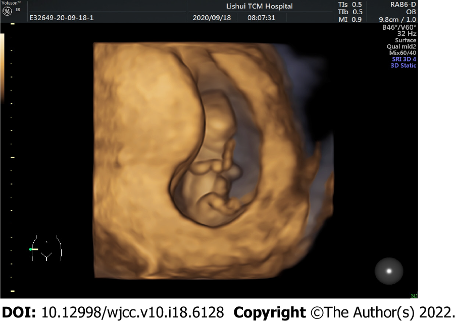
Figure 4 Nuchal translucency examination.
- Citation: Ye JP, Gao Y, Lu LW, Ye YJ. Effectiveness and safety of ultrasound-guided intramuscular lauromacrogol injection combined with hysteroscopy in cervical pregnancy treatment: A case report. World J Clin Cases 2022; 10(18): 6128-6135
- URL: https://www.wjgnet.com/2307-8960/full/v10/i18/6128.htm
- DOI: https://dx.doi.org/10.12998/wjcc.v10.i18.6128


