Copyright
©2013 Baishideng.
World J Clin Cases. May 16, 2013; 1(2): 84-86
Published online May 16, 2013. doi: 10.12998/wjcc.v1.i2.84
Published online May 16, 2013. doi: 10.12998/wjcc.v1.i2.84
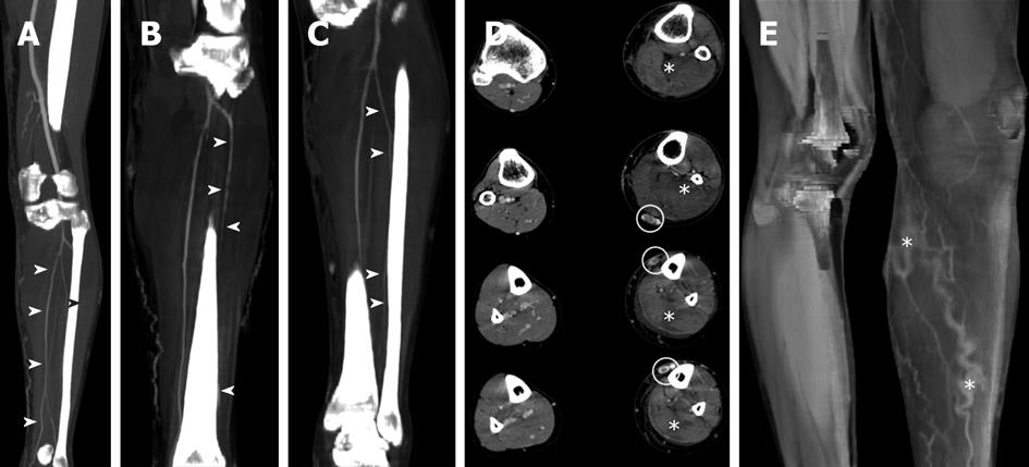
Figure 1 A computed tomography of the lower leg revealed a swollen compartment without vascular lesions and hypertension at the venous end of the capillary beds.
A, B, C: The reconstruction MIP/3D that showed the viability of posterior tibial (A), anterior tibial (B) and interosseal artery (C) (marked by headarrows); D: The swollen muscle compartment (marked by *); E: The presence of hypertension at the venous end of the capillary beds (marked by *).
- Citation: Milone M, Venetucci P, Iervolino S, Taffuri C, Salvatore G, Milone F. A rare case of acute compartment syndrome after saphenectomy. World J Clin Cases 2013; 1(2): 84-86
- URL: https://www.wjgnet.com/2307-8960/full/v1/i2/84.htm
- DOI: https://dx.doi.org/10.12998/wjcc.v1.i2.84


