Copyright
©The Author(s) 2015.
World J Nephrol. Jul 6, 2015; 4(3): 438-443
Published online Jul 6, 2015. doi: 10.5527/wjn.v4.i3.438
Published online Jul 6, 2015. doi: 10.5527/wjn.v4.i3.438
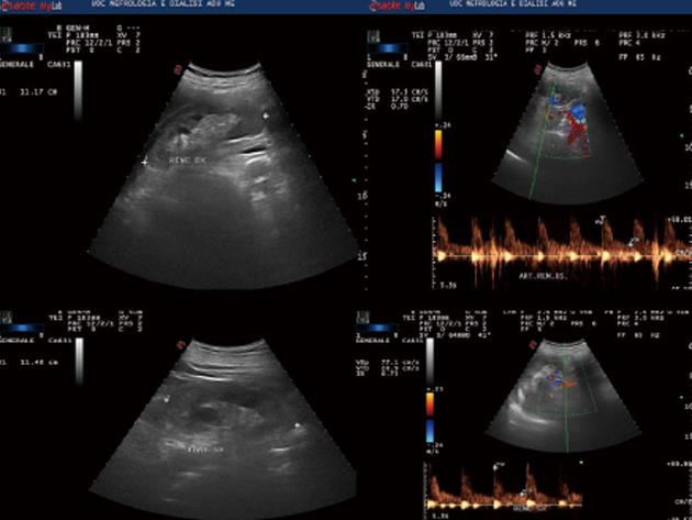
Figure 1 Renal ultrasound.
Figure 2 Chest X-ray.
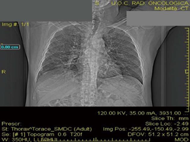
Figure 3 Pulmonary computed tomography.
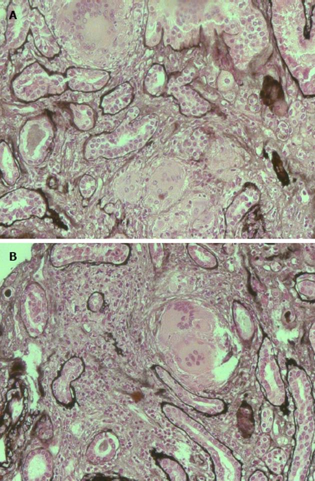
Figure 4 Histological findings.
A: Presence of interstitial multiple granulomas composed of epithelioid cells and moderate lymphocyte and monocyte infiltration (silver stain); B: Multinucleated giant cells (silver stain).
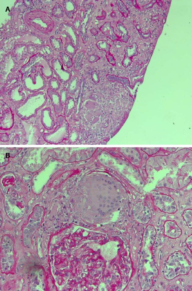
Figure 5 Histological findings.
A: Moderate lymphocyte and monocyte infiltration; B: Multinucleated giant cells [periodic acid schiff (PAS) stain].
- Citation: Lupica R, Buemi M, Campennì A, Trimboli D, Canale V, Cernaro V, Santoro D. Unexpected hypercalcemia in a diabetic patient with kidney disease. World J Nephrol 2015; 4(3): 438-443
- URL: https://www.wjgnet.com/2220-6124/full/v4/i3/438.htm
- DOI: https://dx.doi.org/10.5527/wjn.v4.i3.438


