
















Copyright: ©Author(s) 2026.
World J Crit Care Med. Sep 9, 2026; 15(3): 122632
Published online Sep 9, 2026. doi: 10.5492/wjccm.122632
Published online Sep 9, 2026. doi: 10.5492/wjccm.122632
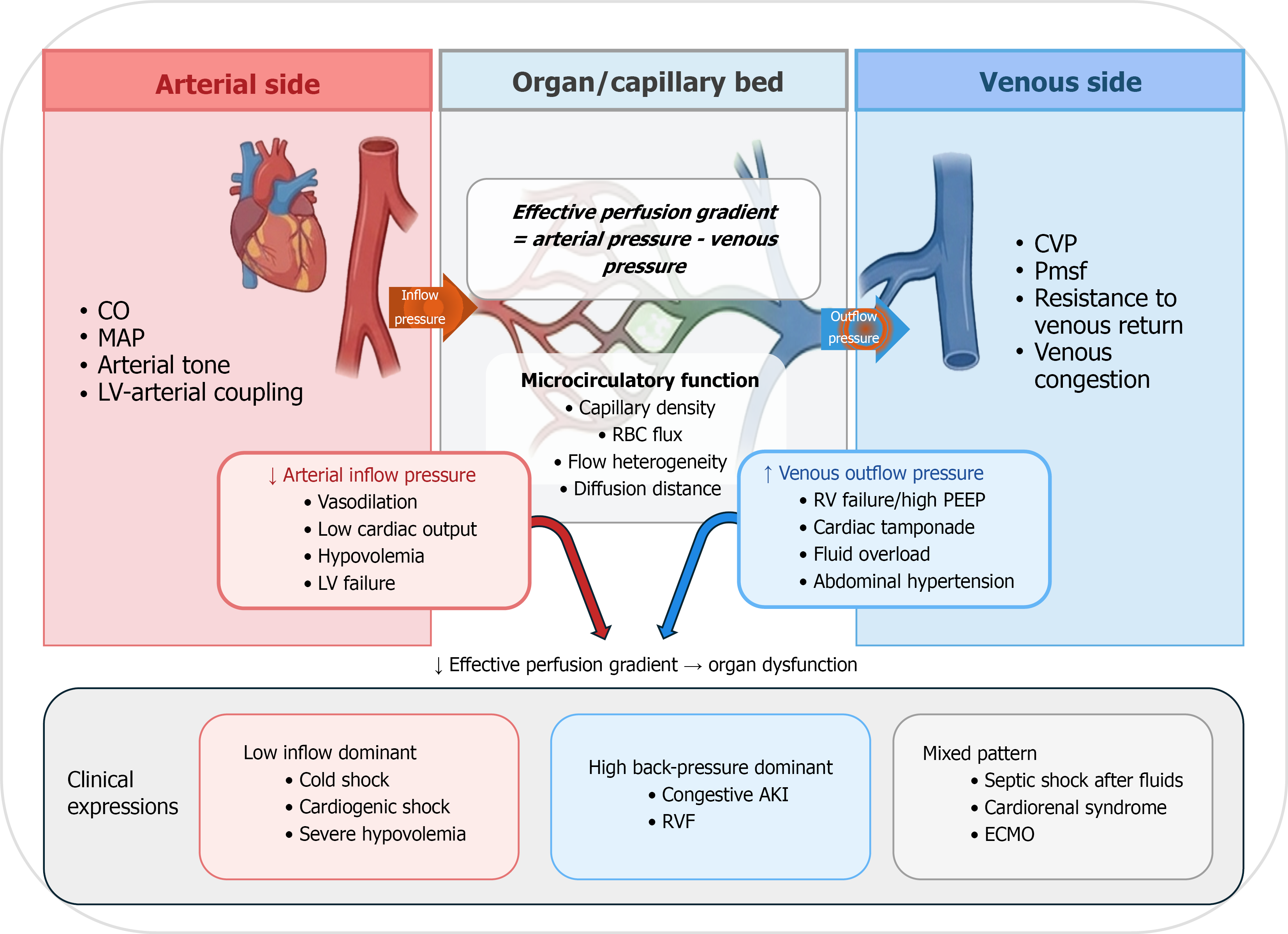
Figure 1 Integrated arterial-venous perfusion gradient model incorporating microcirculatory function.
Schematic representation of the relationship between arterial inflow, venous outflow pressure, and the effective perfusion gradient at the level of the organ capillary bed. The gradient is determined by the difference between arterial and venous pressures and is further modulated by microcirculatory factors including capillary density, red blood cell flux, flow heterogeneity, and diffusion distance. Disturbances in arterial inflow, elevations in venous pressure, or their combination reduce the effective perfusion gradient and contribute to organ dysfunction, with distinct clinical expression patterns. AKI: Acute kidney injury; CO: Cardiac output; CVP: Central venous pressure; ECMO: Extracorporeal membrane oxygenation; LV: Left ventricle; MAP: Mean arterial pressure; PEEP: Positive end-expiratory pressure; RVF: Right ventricular failure; RBC: Red blood cell; RV: Right ventricle; Pmsf: Mean systemic filling pressure.
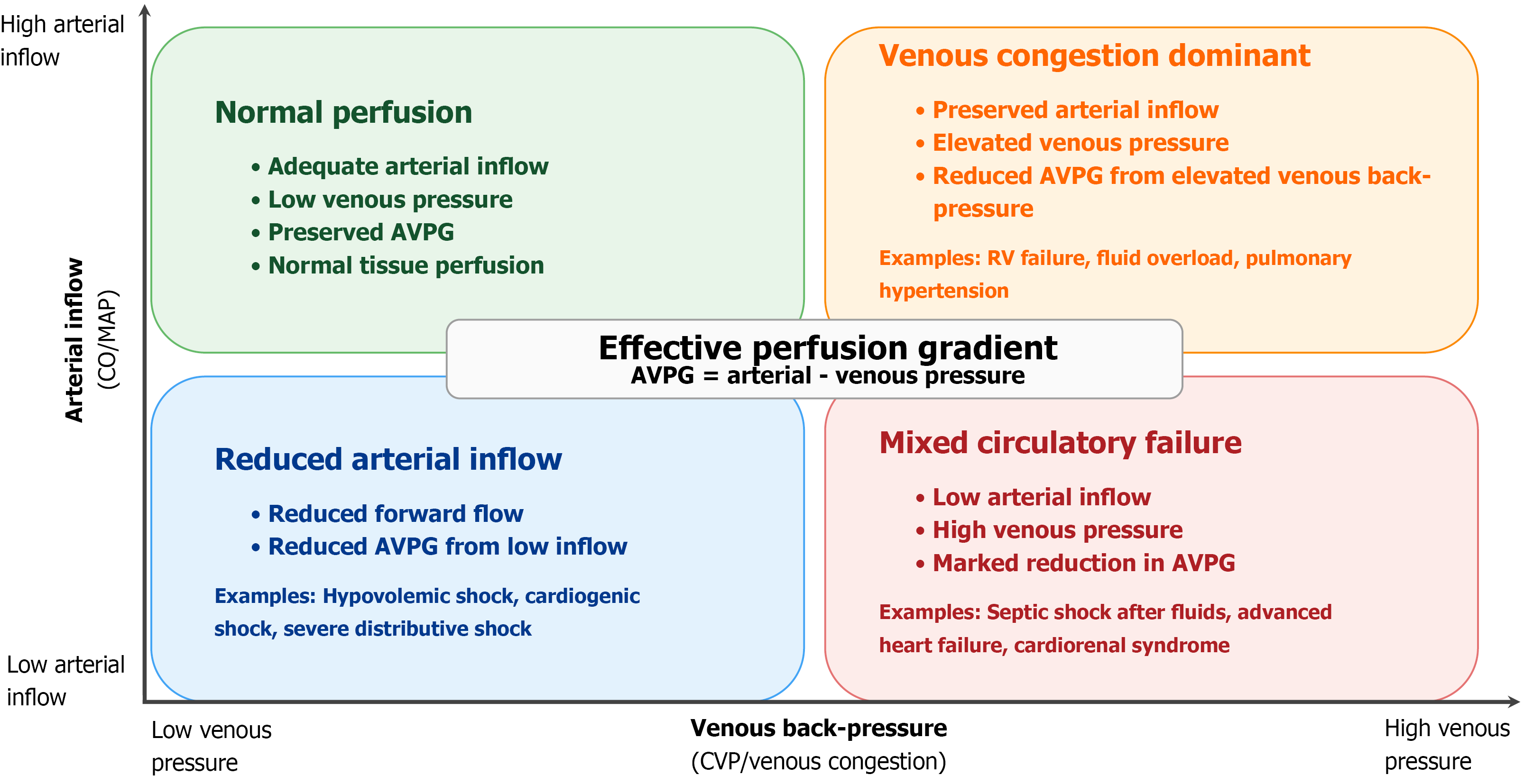
Figure 2 Hemodynamic phenotypes based on the interaction between arterial inflow and venous outflow pressure.
Schematic representation of four hemodynamic patterns defined by arterial inflow and venous pressure, which together determine the effective organ perfusion gradient. Impairment in either or both components leads to reduced tissue perfusion. AVPG: Arterial-venous perfusion gradient; CO: Cardiac output; CVP: Central venous pressure; MAP: Mean arterial pressure.
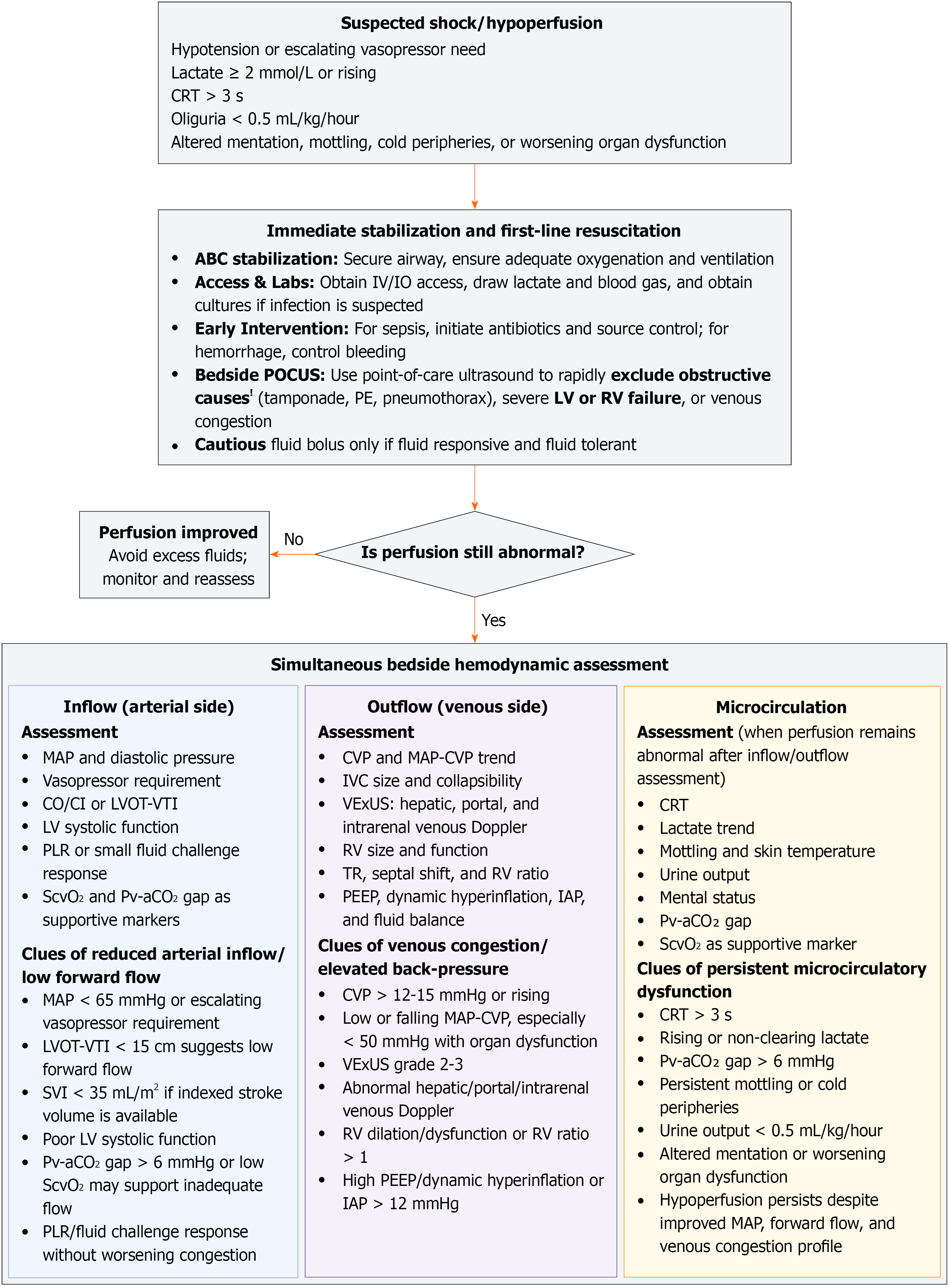
Figure 3 Simultaneous arterial-venous perfusion gradient-based bedside assessment of suspected shock or tissue hypoperfusion.
After initial stabilization, persistent hypoperfusion should be assessed across three domains: Arterial inflow/forward flow, venous outflow/congestion, and microcirculatory perfusion. Suggested values are pragmatic reassessment triggers, not universal treatment thresholds. The aim is to identify whether ongoing hypoperfusion is driven by reduced arterial inflow, venous congestion, combined inflow-congestion failure, or residual microcirculatory dysfunction. ABC: Airway, breathing, circulation; AVPG: Arterial-venous perfusion gradient; CO: Cardiac output; CI: Cardiac index; CRT: Capillary refill time; CVP: Central venous pressure; IAP: Intra-abdominal pressure; IO: Intraosseus; IV: Intravenous; IVC: Inferior vena cava; LVOT-VTI: Left ventricular outflow tract velocity-time integral; MAP: Mean arterial pressure; PE: Pulmonary embolism; PEEP: Positive end-expiratory pressure; PLR: Passive leg raise; POCUS: Point-of-care ultrasound; Pv-aCO₂: Veno-arterial carbon dioxide tension difference; RV: Right ventricle/right ventricular; ScvO₂: Central venous oxygen saturation; SVI: Stroke volume index; VExUS: Venous excess ultrasound score; TR: Tricuspid regurgitation.
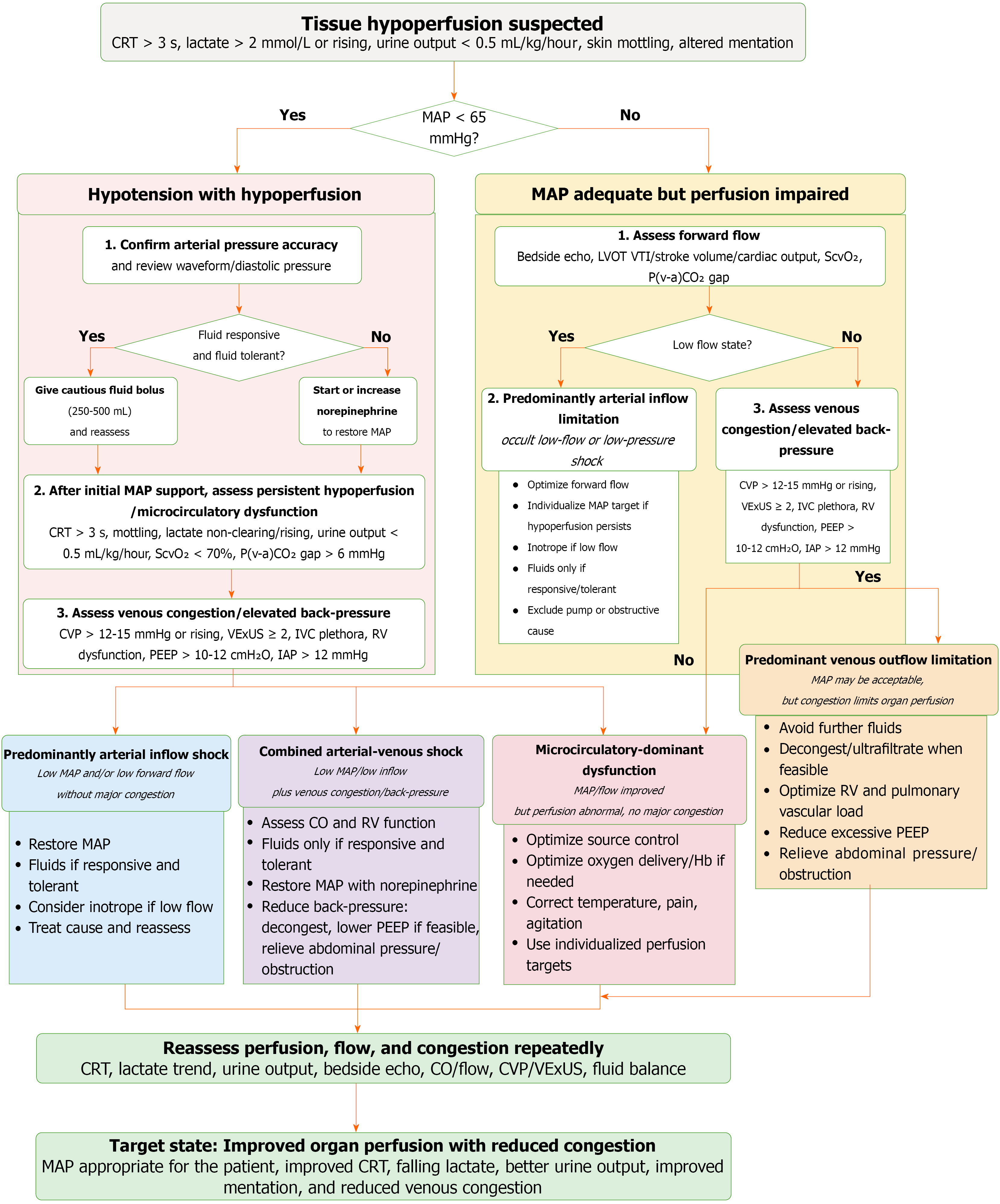
Figure 4 Arterial-venous perfusion gradient-guided bedside pathway for tissue hypoperfusion.
The pathway integrates mean arterial pressure (MAP), forward flow, venous congestion, and tissue perfusion markers to classify suspected hypoperfusion into arterial inflow shock, predominant venous congestion/venous outflow limitation, combined arterial-venous shock, or microcirculatory-dominant dysfunction. Suggested values such as MAP < 65 mmHg, capillary refill time > 3 seconds, lactate > 2 mmol/L or rising, urine output < 0.5 mL/kg/hour, fluid responsiveness, central venous pressure > 12-15 mmHg or rising, venous excess ultrasound score grade 2-3, right ventricle dysfunction, high positive end-expiratory pressure/dynamic hyperinflation, and intra-abdominal pressure > 12 mmHg are pragmatic reassessment triggers rather than universal treatment thresholds. Management is directed toward the dominant phenotype, followed by repeated reassessment of perfusion, flow, congestion, and organ function. The desired endpoint is improved organ perfusion with reduced venous congestion, rather than achievement of a single pressure target. AVPG: Arterial-venous perfusion gradient; CO: Cardiac output; CRT: Capillary refill time; CVP: Central venous pressure; Hb: Hemoglobin; IAP: Intra-abdominal pressure; IVC: Inferior vena cava; LVOT-VTI: Left ventricular outflow tract velocity-time integral; MAP: Mean arterial pressure; PEEP: Positive end-expiratory pressure; P(v-a)CO₂: Veno-arterial carbon dioxide tension difference; RV: Right ventricle/right ventricular; ScvO₂: Central venous oxygen saturation; VExUS: Venous excess ultrasound score.
- Citation: Kataria S, Vinjamuri S, Juneja D, Goel S. Venous dimension of shock: Integrating arterial inflow and venous back-pressure in hemodynamic assessment. World J Crit Care Med 2026; 15(3): 122632
- URL: https://www.wjgnet.com/2220-3141/full/v15/i3/122632.htm
- DOI: https://dx.doi.org/10.5492/wjccm.122632