
















Copyright: ©Author(s) 2026.
World J Crit Care Med. Sep 9, 2026; 15(3): 122632
Published online Sep 9, 2026. doi: 10.5492/wjccm.122632
Published online Sep 9, 2026. doi: 10.5492/wjccm.122632
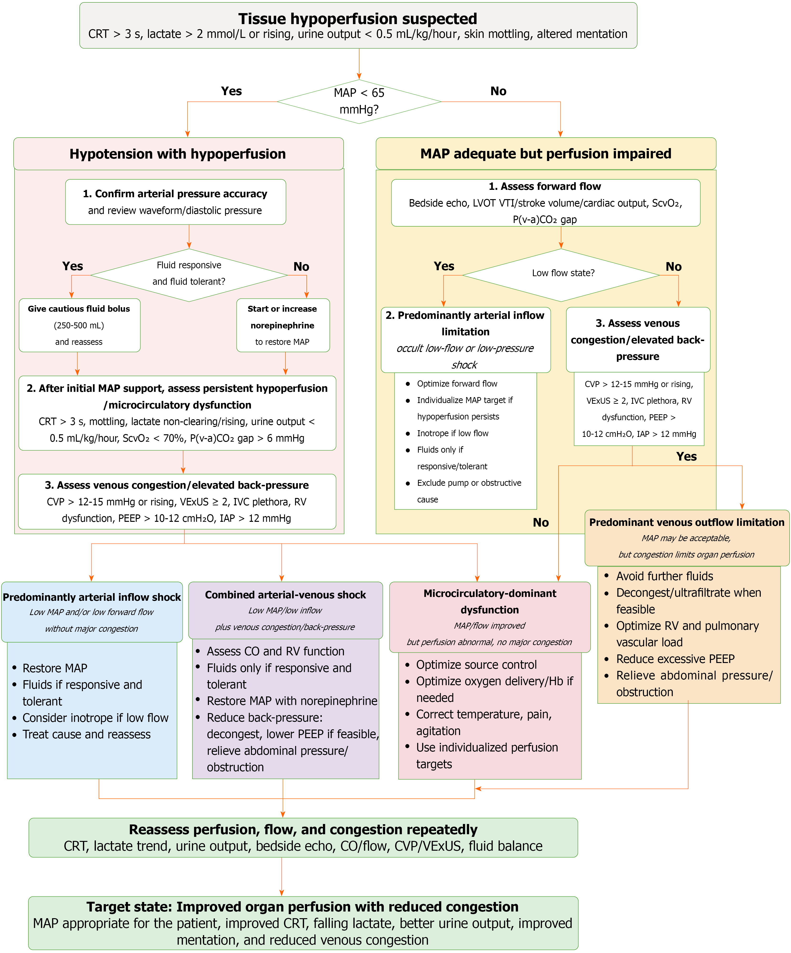
Figure 4 Arterial-venous perfusion gradient-guided bedside pathway for tissue hypoperfusion.
The pathway integrates mean arterial pressure (MAP), forward flow, venous congestion, and tissue perfusion markers to classify suspected hypoperfusion into arterial inflow shock, predominant venous congestion/venous outflow limitation, combined arterial-venous shock, or microcirculatory-dominant dysfunction. Suggested values such as MAP < 65 mmHg, capillary refill time > 3 seconds, lactate > 2 mmol/L or rising, urine output < 0.5 mL/kg/hour, fluid responsiveness, central venous pressure > 12-15 mmHg or rising, venous excess ultrasound score grade 2-3, right ventricle dysfunction, high positive end-expiratory pressure/dynamic hyperinflation, and intra-abdominal pressure > 12 mmHg are pragmatic reassessment triggers rather than universal treatment thresholds. Management is directed toward the dominant phenotype, followed by repeated reassessment of perfusion, flow, congestion, and organ function. The desired endpoint is improved organ perfusion with reduced venous congestion, rather than achievement of a single pressure target. AVPG: Arterial-venous perfusion gradient; CO: Cardiac output; CRT: Capillary refill time; CVP: Central venous pressure; Hb: Hemoglobin; IAP: Intra-abdominal pressure; IVC: Inferior vena cava; LVOT-VTI: Left ventricular outflow tract velocity-time integral; MAP: Mean arterial pressure; PEEP: Positive end-expiratory pressure; P(v-a)CO₂: Veno-arterial carbon dioxide tension difference; RV: Right ventricle/right ventricular; ScvO₂: Central venous oxygen saturation; VExUS: Venous excess ultrasound score.
- Citation: Kataria S, Vinjamuri S, Juneja D, Goel S. Venous dimension of shock: Integrating arterial inflow and venous back-pressure in hemodynamic assessment. World J Crit Care Med 2026; 15(3): 122632
- URL: https://www.wjgnet.com/2220-3141/full/v15/i3/122632.htm
- DOI: https://dx.doi.org/10.5492/wjccm.122632