
















Copyright: ©Author(s) 2026.
World J Crit Care Med. Sep 9, 2026; 15(3): 122062
Published online Sep 9, 2026. doi: 10.5492/wjccm.122062
Published online Sep 9, 2026. doi: 10.5492/wjccm.122062
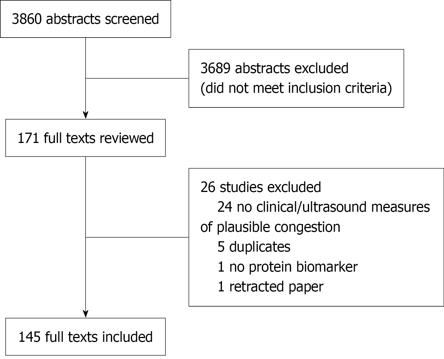
Figure 1 Preferred reporting items for systematic reviews and meta-analysis extension for scoping reviews flow diagram.
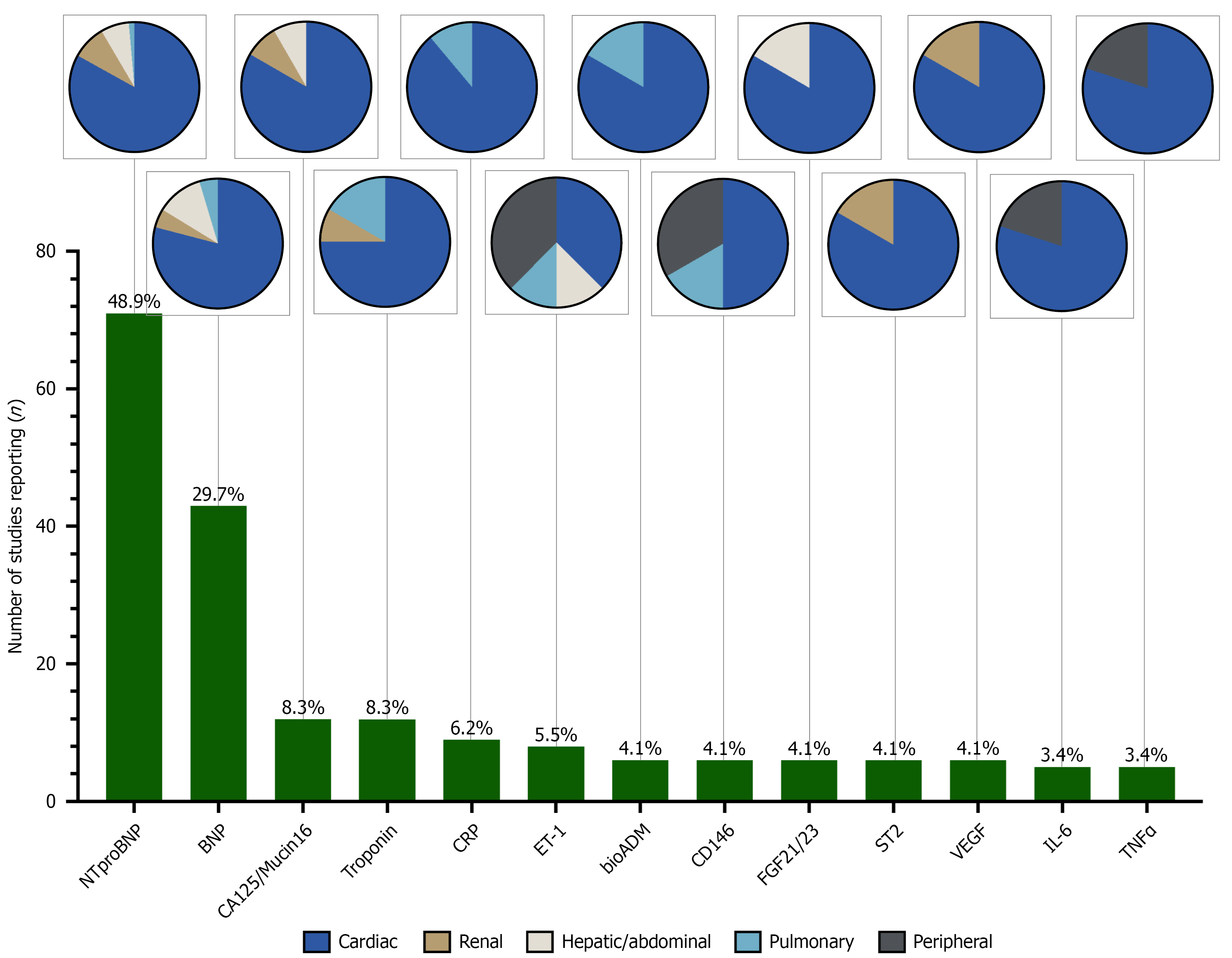
Figure 2 Top circulating markers in studies with measures of venous congestion.
Circulating markers reported by > 3% of studies (5 or more studies) are shown. Cardiovascular and endothelial signaling proteins were the most frequently reported proteins. The bar graph demonstrates the number of studies (Y-axis) that reported clinical associations or findings with the identified proteins (X-axis). The proportion (%) of the 145 studies that report each protein is included above the green bars. The pie charts represent the disease states in which the proteins were quantified (cardiac: Dark blue; pulmonary: Light blue; renal: Beige; hepatic/abdominal: White; peripheral congestion: Grey). A complete list of all reported circulating markers with associations with results or outcomes in studies with measures of venous congestion is shown in Supplementary Table 2. BNP: B-type natriuretic peptide; ET-1: Endothelin 1; VEGF: Vascular endothelial growth factor; CRP: C-reactive protein; IL-6: Interleukin-6; TNF: Tumor necrosis factor; FGF: Fibroblast growth factor.
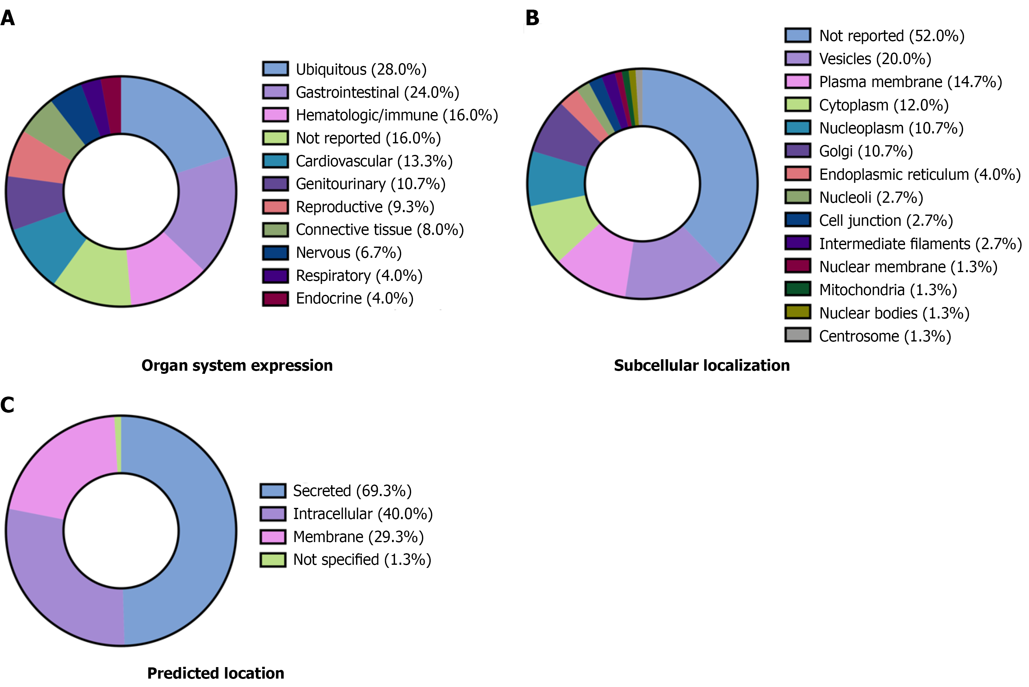
Figure 3 Tissue expression, cellular localization, and predicted location of all identified circulating proteins.
The Human Protein Atlas was used to characterize protein expression across tissues and cells for those circulating proteins that had associations with study results or outcomes in studies with measures of venous congestion (those markers from Supplementary Table 2). Data are presented as a percentage (%) of the 75 unique proteins. Some proteins had multiple localizations. A: Characterization of organ-specific expression; B: Subcellular location based on immunohistochemistry staining; C: Protein localization (secreted, membrane-attached, or intracellular).
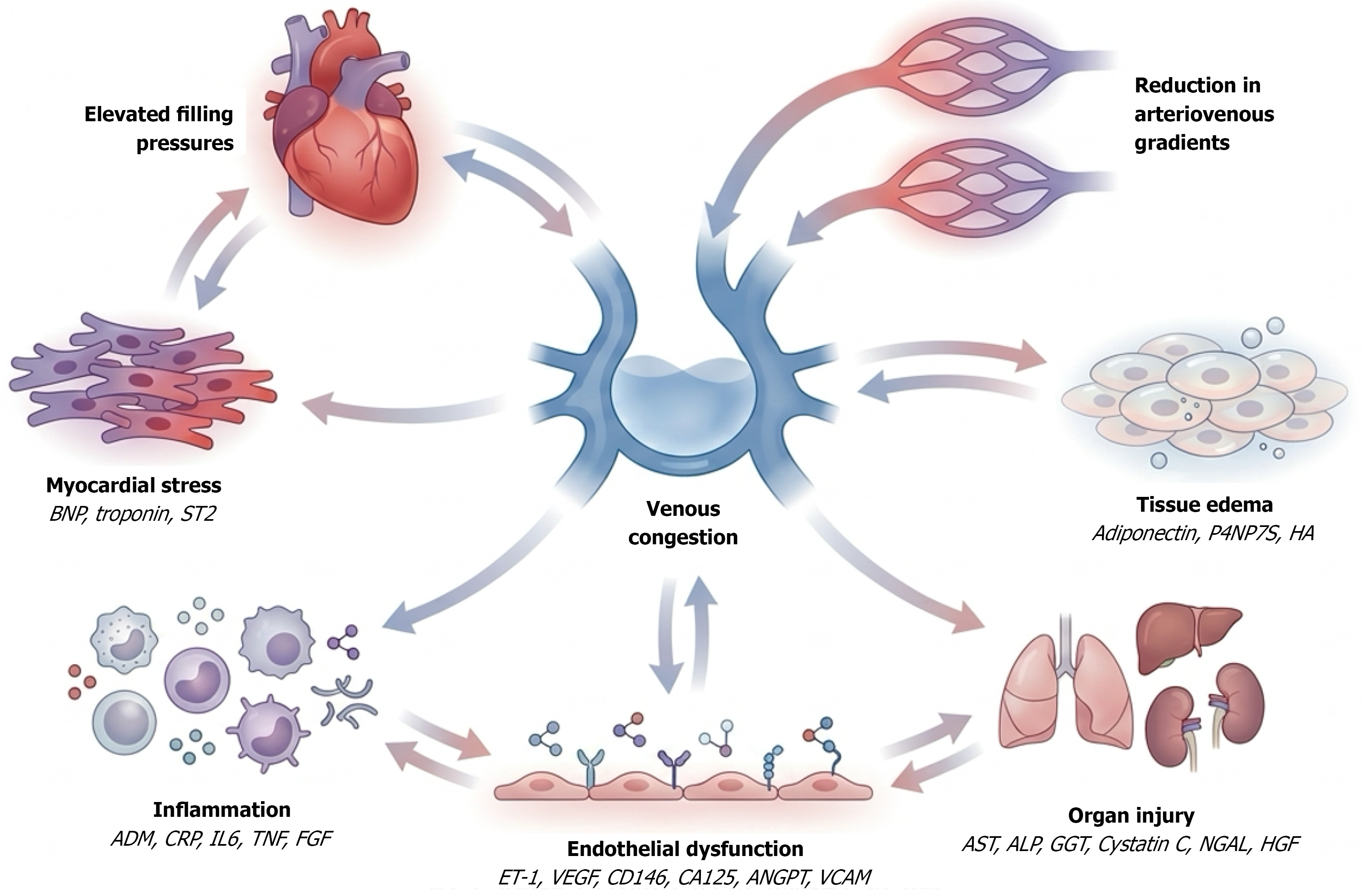
Figure 4 Conceptual model of multisystem molecular dysregulation in venous congestion.
Multiple pathobiologic mechanisms of venous congestion may be associated with the identified candidate circulating protein biomarkers. In venous congestion, elevated filling pressures in the heart may lead to myocardial stress (↑B-type natriuretic peptide, troponin, interleukin 1 receptor-like 1). This may contribute to endothelial dysfunction (↑endothelin 1, vascular endothelial growth factor, CD146, CA125, angiopoietin, vascular cell adhesion molecule 1), promoting vascular permeability and inflammation. Tissue edema (↑adiponectin, 7S domain of collagen type IV and hyaluronic acid) and systemic inflammatory processes (↑adrenomedullin, C-reactive protein, interleukin-6, tumor necrosis factor, fibroblast growth factor) may exacerbate organ dysfunction and, left unresolved, can lead to multiorgan failure involving the lungs, brain, liver, and kidneys (↑aspartate aminotransferase, alkaline phosphatase, gamma glutamyltransferase, Cystatin C, neutrophil gelatinase associated lipocalin, and hepatocyte growth factor). This interconnected network highlights the complex nature of venous congestion and its systemic effects that are yet to be investigated in critical illness. BNP: B-type natriuretic peptide; ST2: Interleukin 1 receptor-like 1; ET-1: Endothelin 1; VEGF: Vascular endothelial growth factor; CA125: Cancer antigen 125; ANGPT: Angiopoietin; VCAM: Vascular cell adhesion molecule 1; ADM: Adrenomedullin; CRP: C-reactive protein; IL-6: Interleukin-6; TNF: Tumor necrosis factor; FGF: Fibroblast growth factor; AST: Aspartate aminotransferase; ALP: Alkaline phosphatase; GGT: Gamma glutamyltransferase; NGAL: Neutrophil gelatinase associated lipocalin; HGF: Hepatocyte growth factor; P4NP7S: 7S domain of collagen type IV; HA: Hyaluronic acid.
- Citation: Van Nynatten LR, Nabieva K, Barber G, Moroniti JJ, Iansavitchene A, Prager R, Slessarev M, Basmaji J, Leligdowicz A. Characterizing the molecular landscape of venous congestion. World J Crit Care Med 2026; 15(3): 122062
- URL: https://www.wjgnet.com/2220-3141/full/v15/i3/122062.htm
- DOI: https://dx.doi.org/10.5492/wjccm.122062