
















Copyright: ©Author(s) 2026.
World J Crit Care Med. Sep 9, 2026; 15(3): 122062
Published online Sep 9, 2026. doi: 10.5492/wjccm.122062
Published online Sep 9, 2026. doi: 10.5492/wjccm.122062
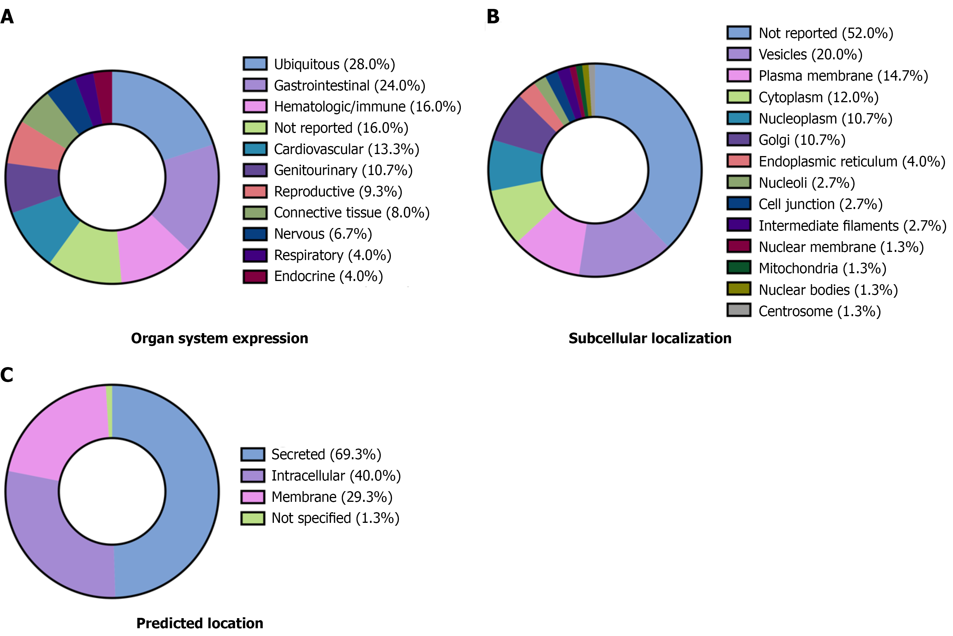
Figure 3 Tissue expression, cellular localization, and predicted location of all identified circulating proteins.
The Human Protein Atlas was used to characterize protein expression across tissues and cells for those circulating proteins that had associations with study results or outcomes in studies with measures of venous congestion (those markers from Supplementary Table 2). Data are presented as a percentage (%) of the 75 unique proteins. Some proteins had multiple localizations. A: Characterization of organ-specific expression; B: Subcellular location based on immunohistochemistry staining; C: Protein localization (secreted, membrane-attached, or intracellular).
- Citation: Van Nynatten LR, Nabieva K, Barber G, Moroniti JJ, Iansavitchene A, Prager R, Slessarev M, Basmaji J, Leligdowicz A. Characterizing the molecular landscape of venous congestion. World J Crit Care Med 2026; 15(3): 122062
- URL: https://www.wjgnet.com/2220-3141/full/v15/i3/122062.htm
- DOI: https://dx.doi.org/10.5492/wjccm.122062