Copyright
©2014 Baishideng Publishing Group Inc.
World J Immunol. Nov 27, 2014; 4(3): 174-184
Published online Nov 27, 2014. doi: 10.5411/wji.v4.i3.174
Published online Nov 27, 2014. doi: 10.5411/wji.v4.i3.174
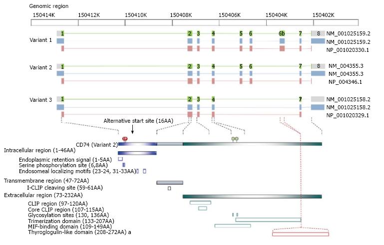
Figure 1 The variants and the corresponding protein structures of cluster of differentiation 74.
The upper panel illustrates the corresponding position among genomic region, NCBI reference sequence number and reference protein accession, exon (green box) and intron (green line) localization of DNA, transcripts (light blue box), and protein (pink box) of the three cluster of differentiation 74 (CD74) variants. The lower panel, CD74 variants contains three regions including intracellular, transmembrane and extracellular regions with the indicated functional domains and identified residues for post-translational modification. Variant 2 transcripts two isoforms, p33 and p35 caused by the alternative start site. Variant 1 transcripts two isoforms, p41 and p43, with an exon 6b-encoded thyroglobulin type I domain to interact with cathepsin. Variant 3 lacks exon 5, 6, and 6b, which translates truncated trimerization domain and truncated macrophage migration inhibitory factor (MIF)-binding domain and remains only the CLIP region to function as major histocompatibility complex class II mask. The amino acid residues refer to human variant 2 (p35).
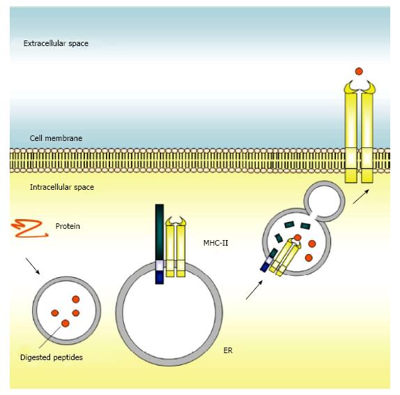
Figure 2 The canonical function of cluster of differentiation 74 in the immune system.
Cluster of differentiation 74 (CD74) is present on endoplasmic reticulum (ER) where it can interact with major histocompatibility complex class II (MHC class II) and contribute to antigen presentation. Once synthesized, CD74 self-assembles as a trimer and serves as a scaffold onto which nascent MHC class II assemble. After trafficking to the late endosome, CD74 is cleaved into a small peptide, CLIP, to block the peptide binding cleft of MHC class II, prevent premature binding of antigenic peptides, and direct the MHC class II complex to the endosomal pathway. The MHC class II molecules with bound antigenic peptides are then exported to the surface of the antigen presenting cell for presentation of foreign peptides to CD4+ T cells.
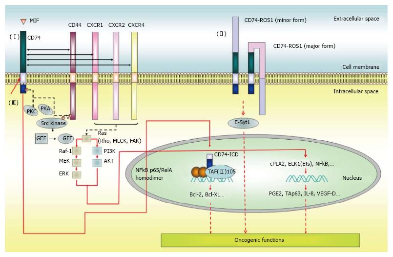
Figure 3 The function of cluster of differentiation 74 in the cancer development.
(I) Membrane associated-CD74 is involved in modulating the expression of a variety of genes which involved cell proliferation, invasion and survival through interacting with CD44, CXCR1, CXCR2 or CXCR4 followed by activation of the signaling cascades in a MIF-dependent manner. (II) After MIF stimulation, CD74 releases its intracellular domain, CD74-ICD. The CD74-ICD translocates from cytoplasm into nucleus and functions as a transcription modulator. (III) The CD74-ROS1 fusion protein with one (minor form) or two (major form) transmembrane regions and one kinase domain, promotes novel invasiveness pathway through the phosphorylation of the extended synaptotagmin-like protein, E-Syt1. MIF: Macrophage migration inhibitory factor; CXCR: Chemokine (C-X-C motif) receptor; PKA: Protein kinase A; PKC: Protein kinase C; GEF: Guanine nucleotide exchange factor; Ras: Ras oncogene; Rho: Ras homolog family member; MLCK: Myosin light chain kinase; FAK: Focal adhesion kinase; Raf-1: RAF proto-oncogene serine/threonine-protein kinase; MEK: Mitogen-activated protein kinase kinase; ERK: Extracellular signal-regulated kinase; PI3K: Phosphoinositide 3-kinase; NF-κB: Nuclear factor-kappaB; TAF(II)105: Transcription initiation factor TFIID 105 kDa subunit; ROS-1: C-ros oncogene 1; E-Syt1: Extended synaptotagmin-like protein 1; Bcl-2: B-cell lymphoma 2; Bcl-XL: B-cell lymphoma-extra large; cPLA2: Cytosolic phospholipase A2; ELK1: Member of ETS oncogene family; Ets: V-ets erythroblastosis virus E26 oncogene; PGE2: Prostaglandin E2; Tap63: Tumor protein p63; IL-8: Interleukin 8; VEGF-D: Vascular endothelial growth factor D.
- Citation: Liu YH, Lin JY. Recent advances of cluster of differentiation 74 in cancer. World J Immunol 2014; 4(3): 174-184
- URL: https://www.wjgnet.com/2219-2824/full/v4/i3/174.htm
- DOI: https://dx.doi.org/10.5411/wji.v4.i3.174


