Copyright
©The Author(s) 2020.
World J Clin Pediatr. Sep 19, 2020; 9(2): 17-28
Published online Sep 19, 2020. doi: 10.5409/wjcp.v9.i2.17
Published online Sep 19, 2020. doi: 10.5409/wjcp.v9.i2.17
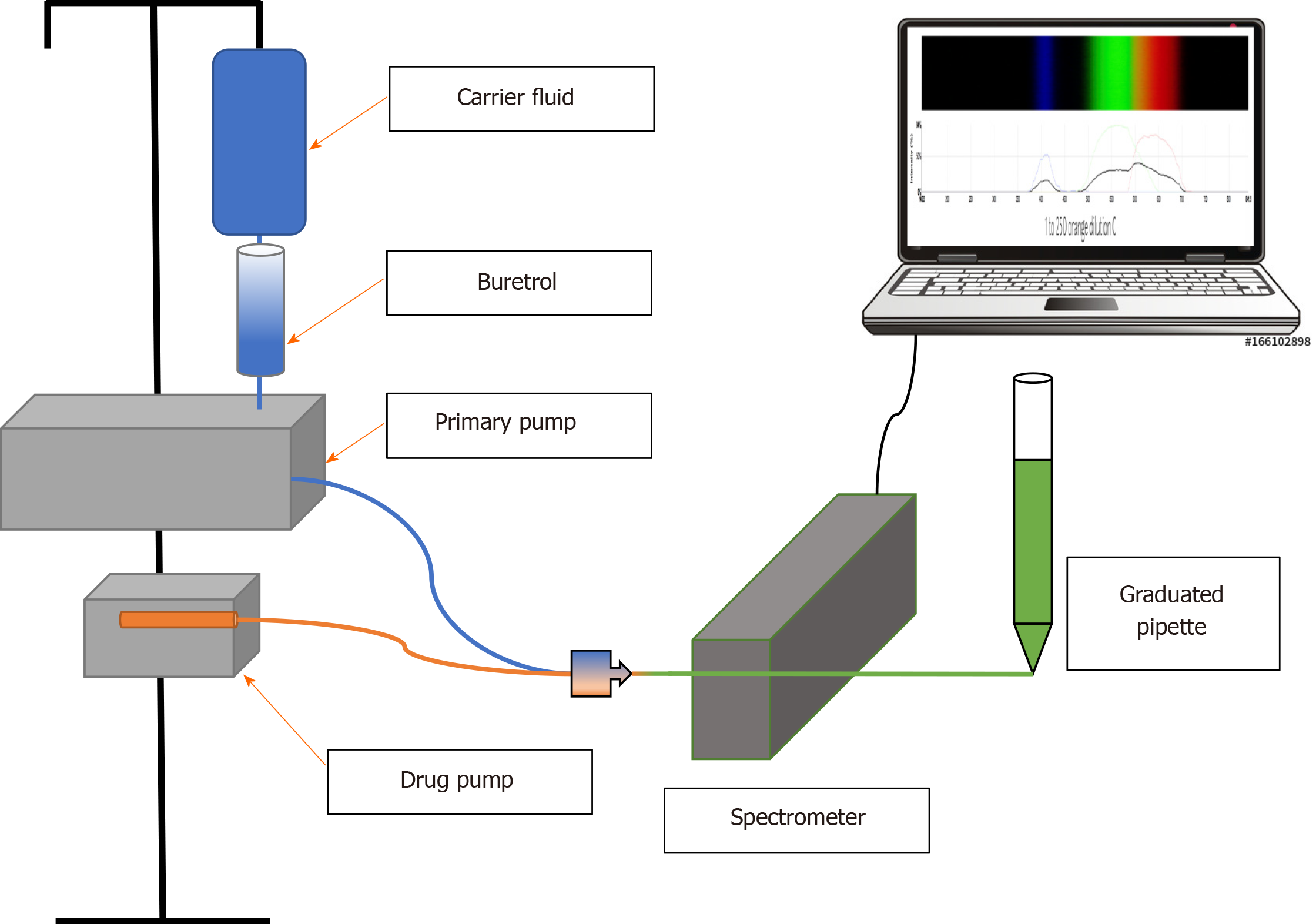
Figure 1 Experimental set-up.
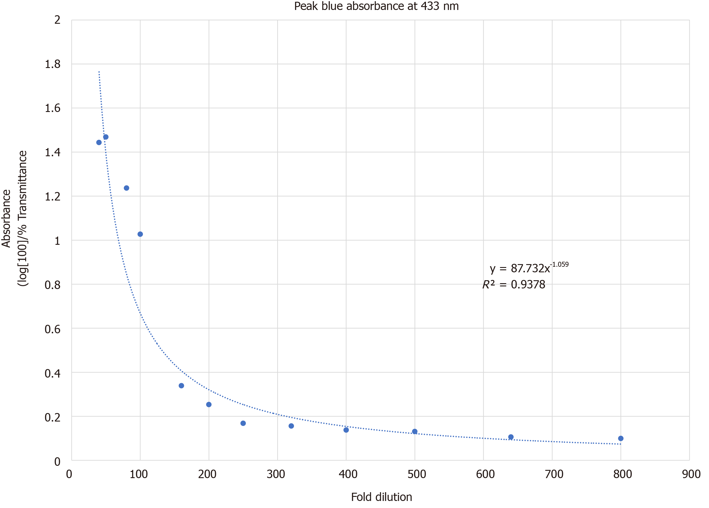
Figure 2 433 nm absorbance vs fold dilution of orange drug in blue carrier fluid.
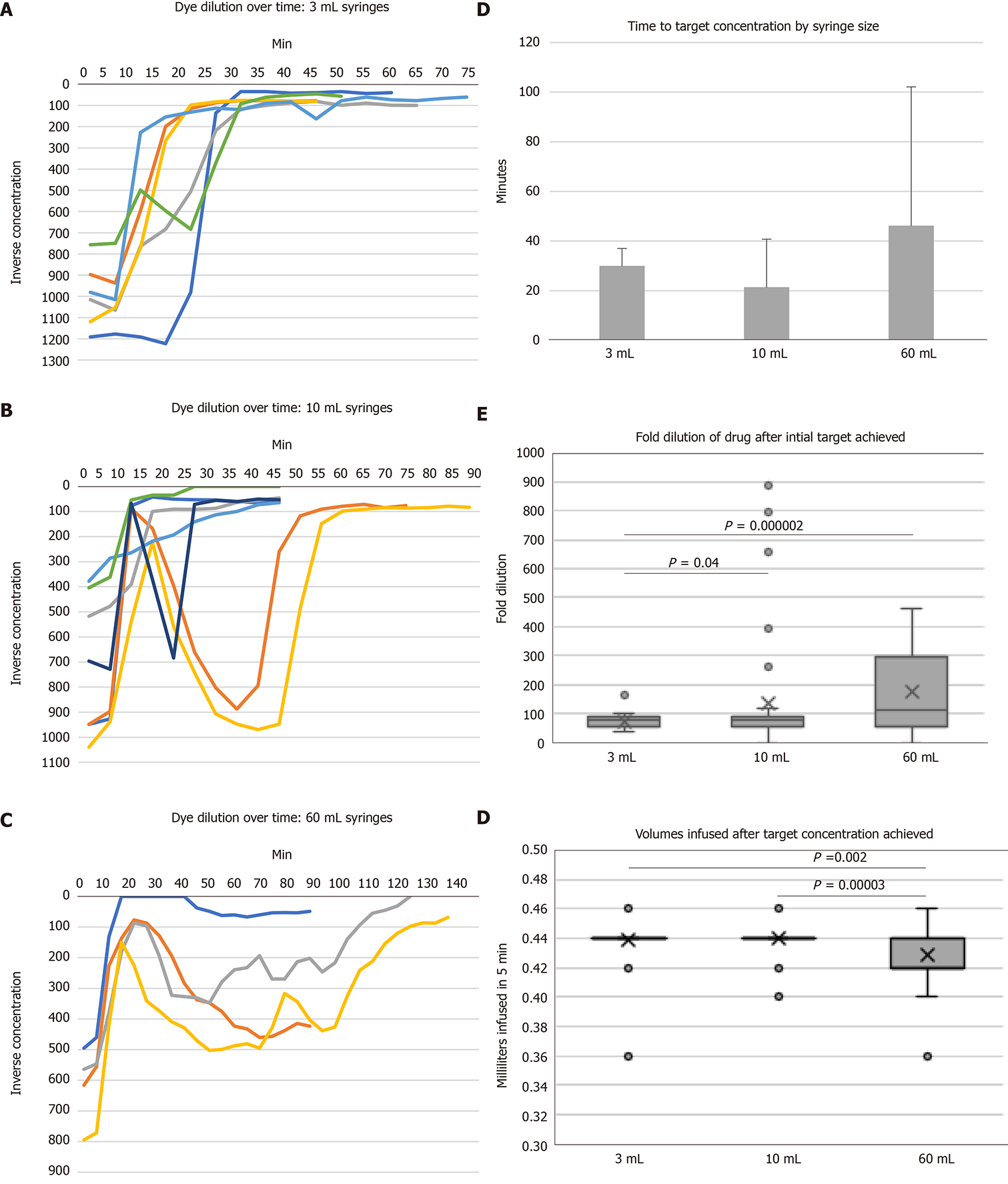
Figure 3 Low flow drug delivery at 0.
2 mL/h with carrier fluid at 5 mL/h. A-C: Orange drug dilution over time; D: Time to target orange drug concentration of 100-fold dilution or less (no significant differences, P = 0.42). Average minutes shown with standard deviation error bars; E and F: Box and whiskers plots with outliers of (E) measured dye dilutions after target concentration achieved (ANOVA P = 0.0067) and (F) volumes infused per 5 min interval after target concentration achieved (ANOVA P = 0.00006).
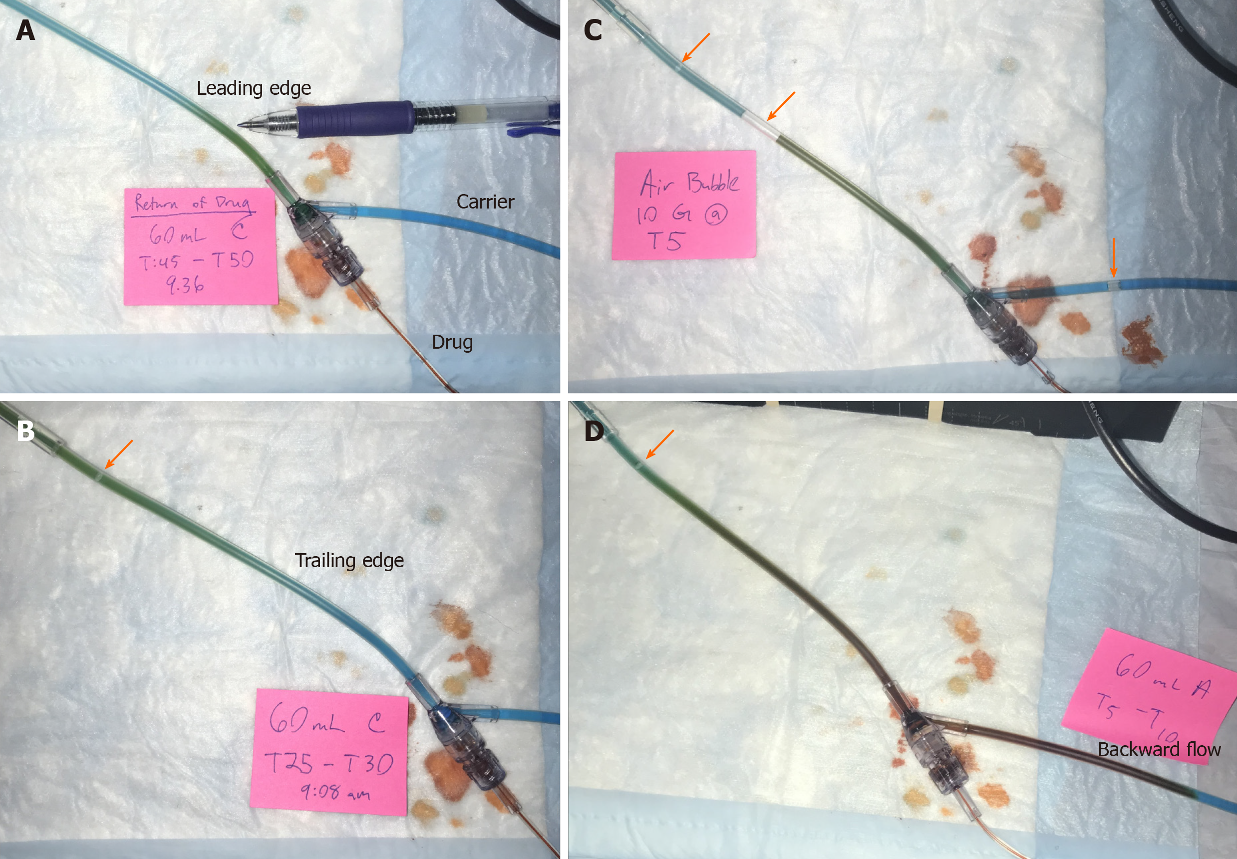
Figure 4 A leading edge of visible color change in the infusion tubing was observed as orange drug traversed the tubing.
A: Green leading edge of admixed orange drug and BL carrier fluid seen after drug under-infusion during a 60 mL syringe trial; B: Trailing edge of green admixed drug and carrier solution during an under-infusion event with a 60 mL syringe, with BL carrier fluid proximally; C: Air bubbles introduced from the carrier line into the infusion set (arrows), which are also seen in (B) and (D); and D: Backward flow of orange drug into the proximal BL carrier fluid line in a 60-mL syringe trial. Note the orange color in the backward fluid column, suggesting lack of drug-carrier fluid mixing, as would be indicated by a transition to green color.
- Citation: Madson ZC, Vangala S, Sund GT, Lin JA. Does carrier fluid reduce low flow drug infusion error from syringe size? World J Clin Pediatr 2020; 9(2): 17-28
- URL: https://www.wjgnet.com/2219-2808/full/v9/i2/17.htm
- DOI: https://dx.doi.org/10.5409/wjcp.v9.i2.17


