Copyright
©The Author(s) 2015.
World J Stomatol. Feb 20, 2015; 4(1): 29-34
Published online Feb 20, 2015. doi: 10.5321/wjs.v4.i1.29
Published online Feb 20, 2015. doi: 10.5321/wjs.v4.i1.29
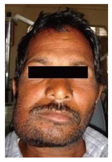
Figure 1 Diffuse swelling on lower right side of face.
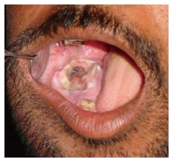
Figure 2 Swelling in molar region with marks of indentation on overlying mucosa.
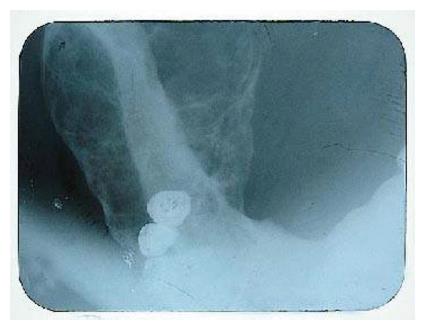
Figure 3 Mandibular lateral occlusal view showing predominantly buccal and lingual expansion with thinning of cortical plates.
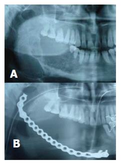
Figure 4 Cropped orthopantomograph showing (A) a multilocular lesion in body and ramus of mandible on right side (B) a surgical defect and radioopaque image of reconstruction plate.
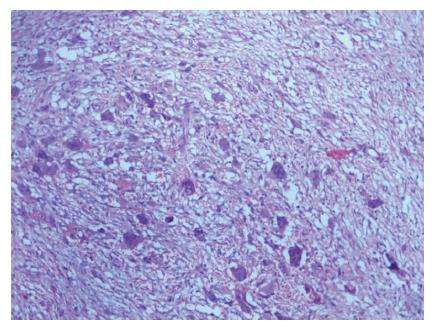
Figure 5 Photomicrograph showing multinucleated giant cells and spindle cells (10 ×).
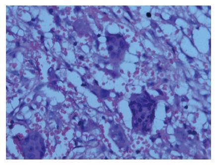
Figure 6 Photomicrograph showing multinucleated giant cells (40 ×).
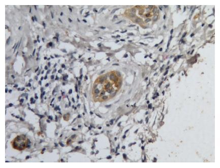
Figure 7 Immunohistochemical expresson of cytokeratin in giant cells.
- Citation: Dangore-Khasbage S, Degwekar SS, Bhowate RR, Hande AH, Lohe VK. Unusual aggressive behavior of central giant cell granuloma following tooth extraction. World J Stomatol 2015; 4(1): 29-34
- URL: https://www.wjgnet.com/2218-6263/full/v4/i1/29.htm
- DOI: https://dx.doi.org/10.5321/wjs.v4.i1.29


