Copyright
©2013 Baishideng.
World J Otorhinolaryngol. Aug 28, 2013; 3(3): 100-107
Published online Aug 28, 2013. doi: 10.5319/wjo.v3.i3.100
Published online Aug 28, 2013. doi: 10.5319/wjo.v3.i3.100
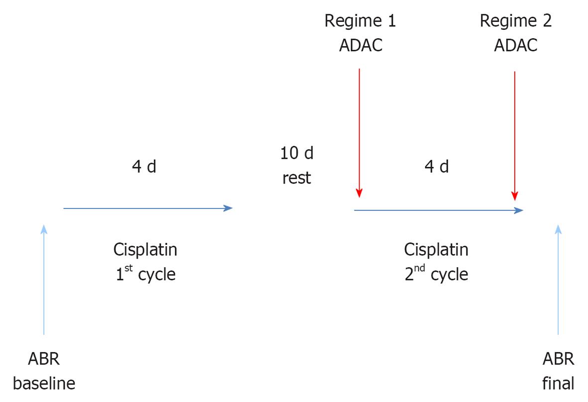
Figure 1 Study design.
Cisplatin injections (1 mg/kg ip) were given twice daily in two cycles separated by 10 d of rest, and adenosine amine congener (ADAC) (100 μg/kg ip) was administered as five daily injections at 24 h intervals. ADAC treatment was administered along with the second cisplatin cycle (Regime 1) or immediately after completion of both cycles (Regime 2).
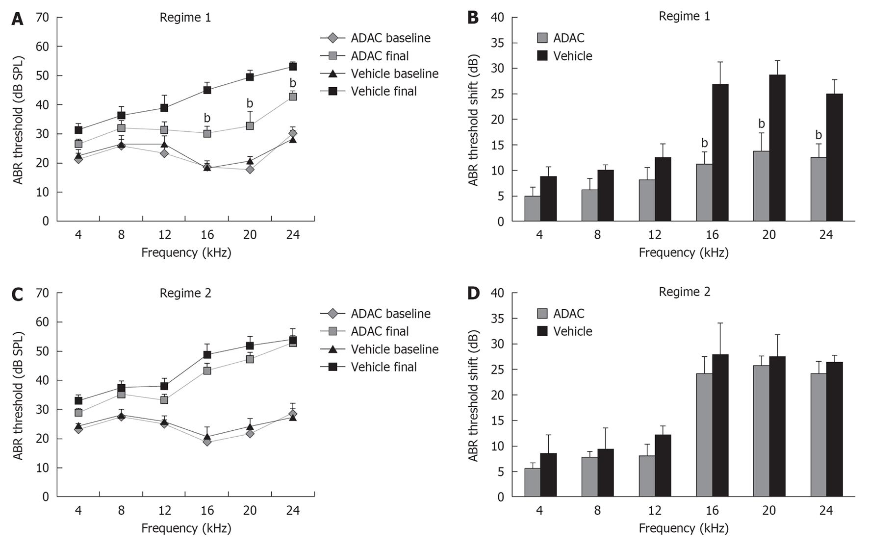
Figure 2 The effect of adenosine amine congener on cisplatin-induced auditory brainstem responses threshold shifts.
A: Auditory brainstem responses (ABR) thresholds before (baseline) and 7 d after cisplatin administration (final). Adenosine amine congener (ADAC) was co-applied with cisplatin during the second cycle (Regime 1); B: ADAC reduced ABR threshold shifts when administered concomitantly with the second cisplatin cycle; C: ABR thresholds before (baseline) and 7 d after cisplatin administration (final). ADAC was administered after the completion of cisplatin treatment (Regime 2); D: ADAC had no effect on cisplatin-induced threshold shifts when applied after the completion of cisplatin treatment. In the control group, injections of the vehicle solution were administered at the same intervals as ADAC. ABRs were measured in response to tone pips (4-24 kHz). Data are expressed as mean ± SE (n = 8). bP < 0.01 vs control group, one-way analysis of variance.
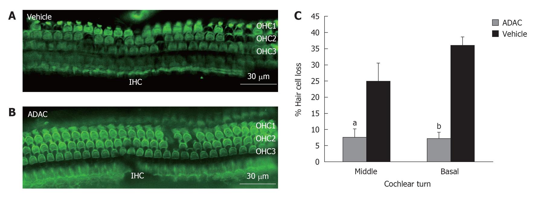
Figure 3 The effect of adenosine amine congener on hair cell loss in the rat cochleae exposed to cisplatin (Regime 1).
A: The surface preparation of the middle turn organ of Corti in the vehicle-treated cochlea; B: The middle turn organ of Corti in the adenosine amine congener (ADAC)-treated cochlea; C: Percentage of hair cell loss in the cochleae exposed to cisplatin treated with ADAC or drug vehicle solution. Data presented as mean ± SE (n = 8). aP < 0.05, bP < 0.01 vs control grou, unpaired t test. IHC: Inner hair cells; OHC: Outer hair cells.
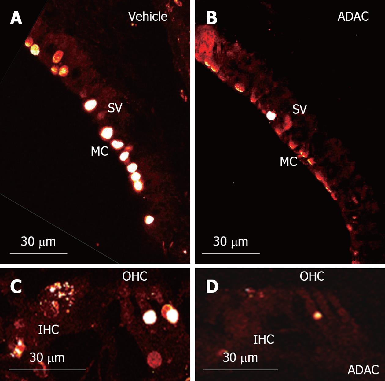
Figure 4 Transferase mediated dUTP nick end labelling staining in the rat cochleae exposed to cisplatin.
A: Apoptotic marginal cells (MC) of the stria vascularis (SV) in the control vehicle-treated cochlea; B: Reduced number of apoptotic marginal cells in the adenosine amine congener (ADAC)-treated cochlea; C: Terminal deoxynucleotidyl transferase mediated dUTP nick end labeling assay staining in the organ of Corti of the control vehicle-treated cochlea; D: Reduced apoptosis in the organ of Corti of the ADAC-treated cochlea. Images are single optical sections of the middle turn. IHC: Inner hair cells; OHC: Outer hair cells.
- Citation: Gunewardene N, Guo CX, Wong AC, Thorne PR, Vlajkovic SM. Adenosine amine congener ameliorates cisplatin-induced hearing loss. World J Otorhinolaryngol 2013; 3(3): 100-107
- URL: https://www.wjgnet.com/2218-6247/full/v3/i3/100.htm
- DOI: https://dx.doi.org/10.5319/wjo.v3.i3.100


