Copyright
©2013 Baishideng.
World J Otorhinolaryngol. Aug 28, 2013; 3(3): 100-107
Published online Aug 28, 2013. doi: 10.5319/wjo.v3.i3.100
Published online Aug 28, 2013. doi: 10.5319/wjo.v3.i3.100
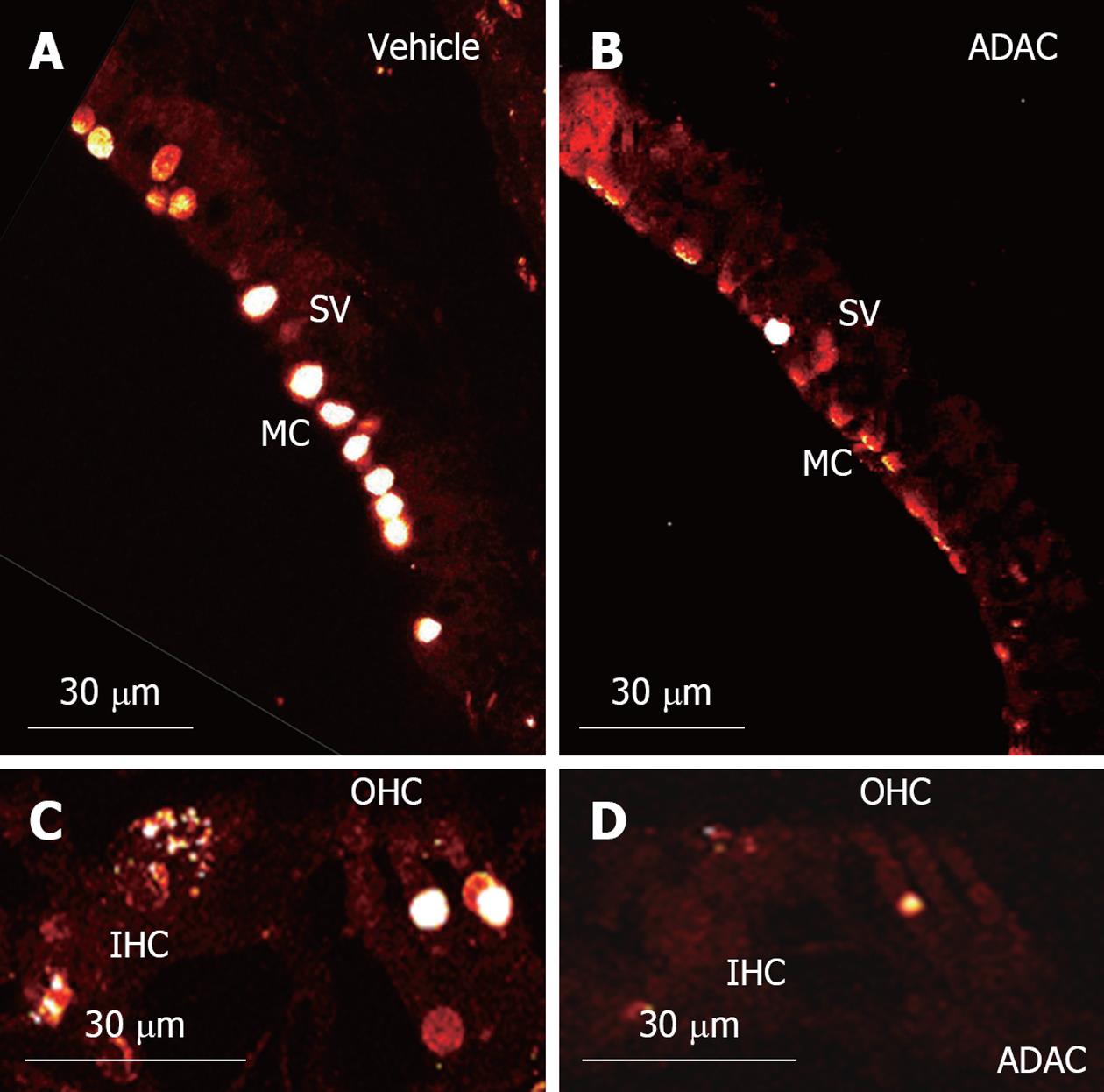
Figure 4 Transferase mediated dUTP nick end labelling staining in the rat cochleae exposed to cisplatin.
A: Apoptotic marginal cells (MC) of the stria vascularis (SV) in the control vehicle-treated cochlea; B: Reduced number of apoptotic marginal cells in the adenosine amine congener (ADAC)-treated cochlea; C: Terminal deoxynucleotidyl transferase mediated dUTP nick end labeling assay staining in the organ of Corti of the control vehicle-treated cochlea; D: Reduced apoptosis in the organ of Corti of the ADAC-treated cochlea. Images are single optical sections of the middle turn. IHC: Inner hair cells; OHC: Outer hair cells.
- Citation: Gunewardene N, Guo CX, Wong AC, Thorne PR, Vlajkovic SM. Adenosine amine congener ameliorates cisplatin-induced hearing loss. World J Otorhinolaryngol 2013; 3(3): 100-107
- URL: https://www.wjgnet.com/2218-6247/full/v3/i3/100.htm
- DOI: https://dx.doi.org/10.5319/wjo.v3.i3.100


