
















Copyright: ©Author(s) 2026.
World J Ophthalmol. Sep 12, 2026; 10(1): 119850
Published online Sep 12, 2026. doi: 10.5318/wjo.119850
Published online Sep 12, 2026. doi: 10.5318/wjo.119850
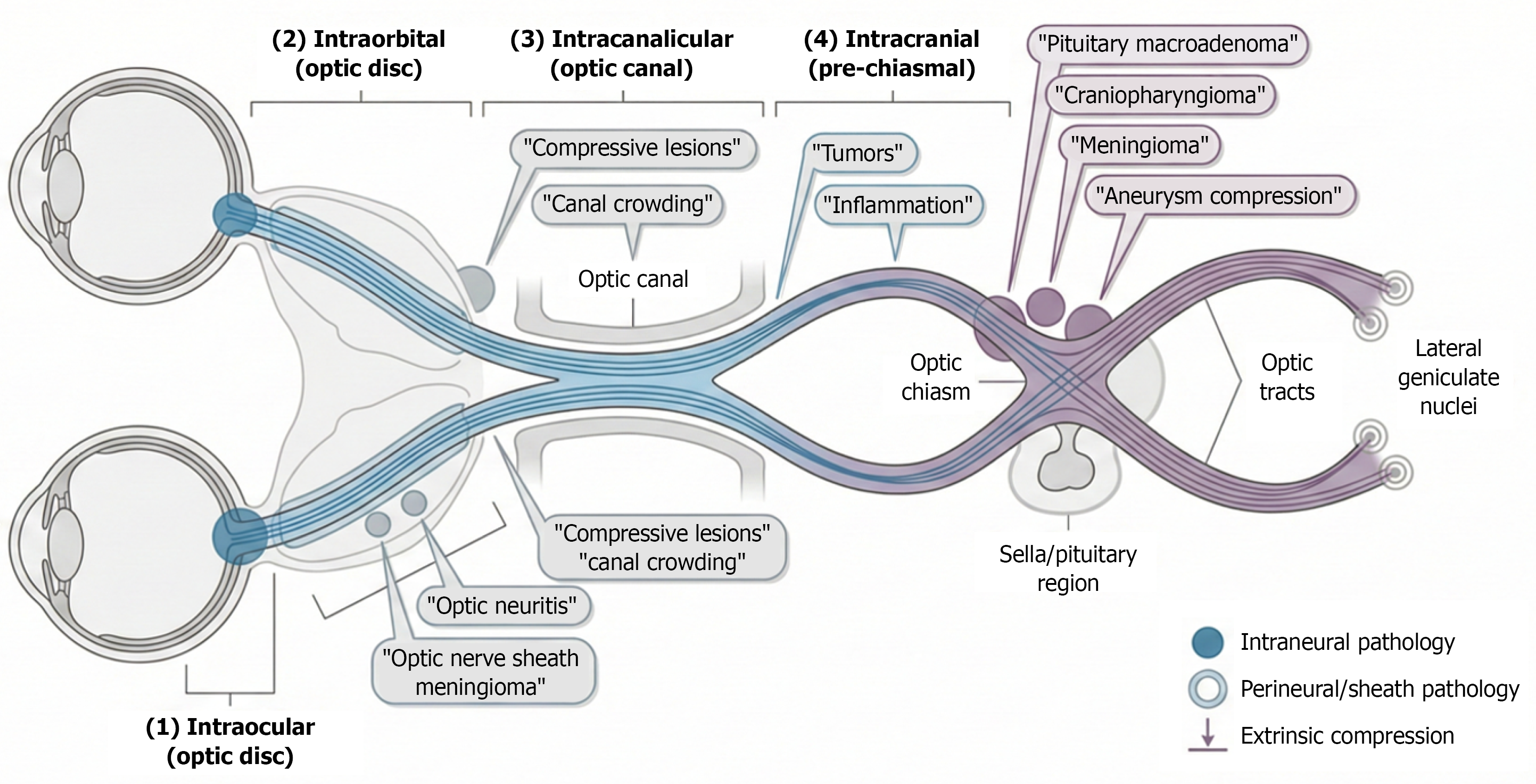
Figure 1 Magnetic resonance imaging-oriented roadmap of the anterior visual pathway.
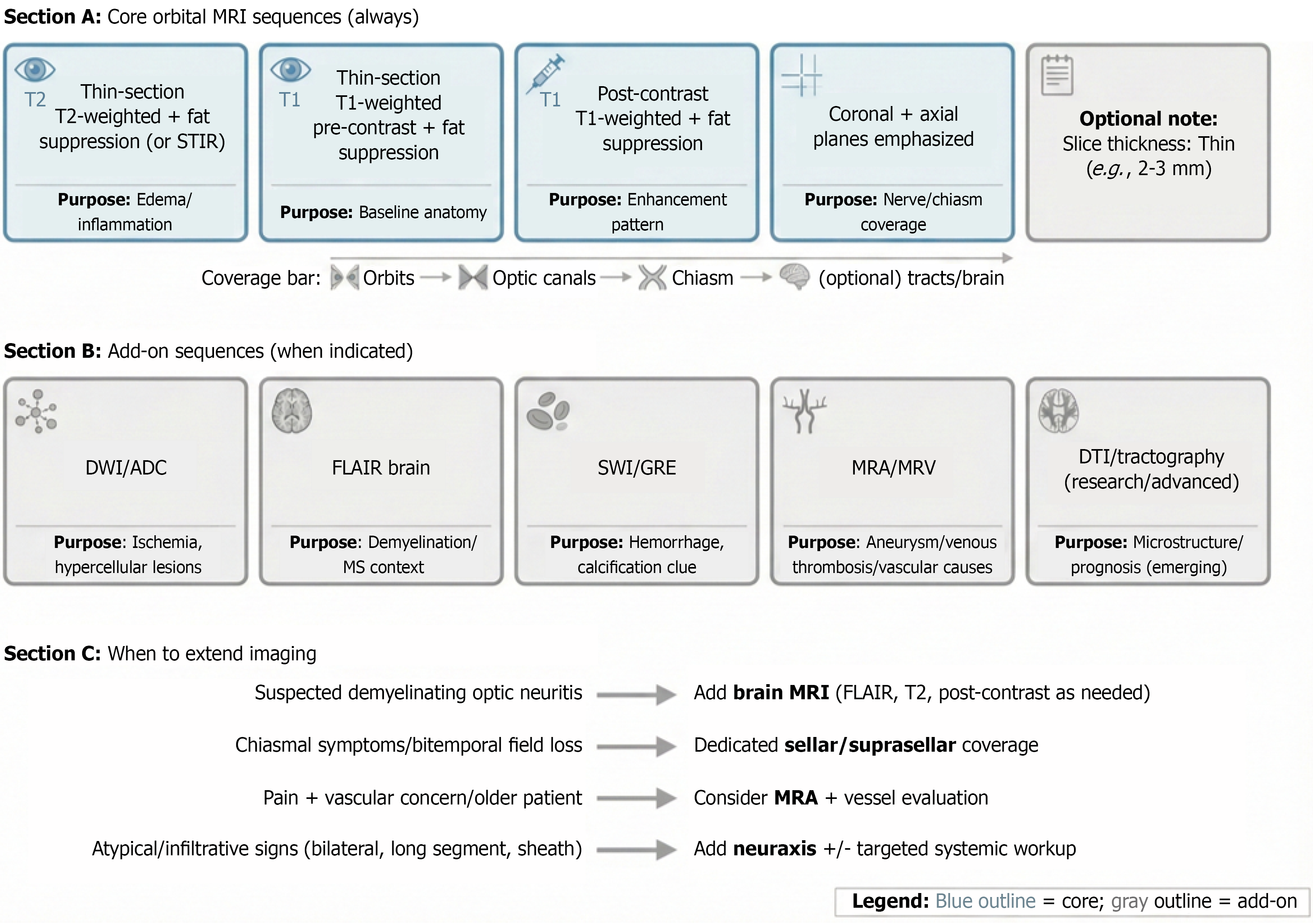
Figure 2 Practical magnetic resonance imaging protocol for optic nerve and chiasmal evaluation.
ADC: Apparent diffusion coefficient; DTI: Diffusion tensor imaging; DWI: Diffusion-weighted imaging; FLAIR: Fluid attenuated inversion recovery; GRE: Gradient echo; MRA: Magnetic resonance angiography; MRI: Magnetic resonance imaging; MRV: Magnetic resonance venography; STIR: Short TI inversion recovery; SWI: Susceptibility-weighted imaging.
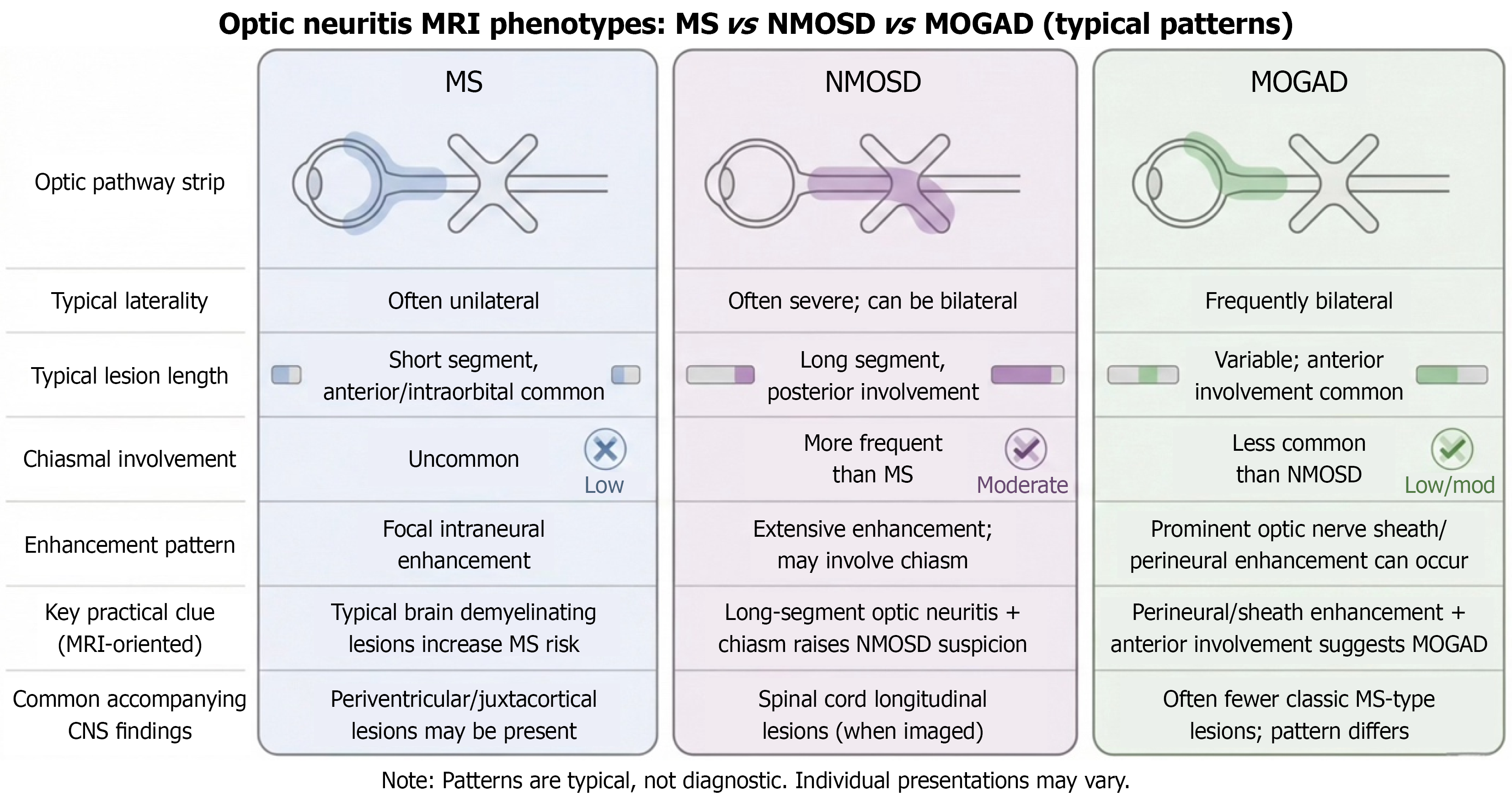
Figure 3 Comparative magnetic resonance imaging phenotypes of multiple sclerosis-associated, neuromyelitis optica spectrum disorder-associated, and myelin oligodendrocyte glycoprotein antibody-associated disease-associated optic neuritis.
MOGAD: Myelin oligodendrocyte glycoprotein antibody-associated disease; MRI: Magnetic resonance imaging; MS: Multiple sclerosis; NMOSD: Neuromyelitis optica spectrum disorder.
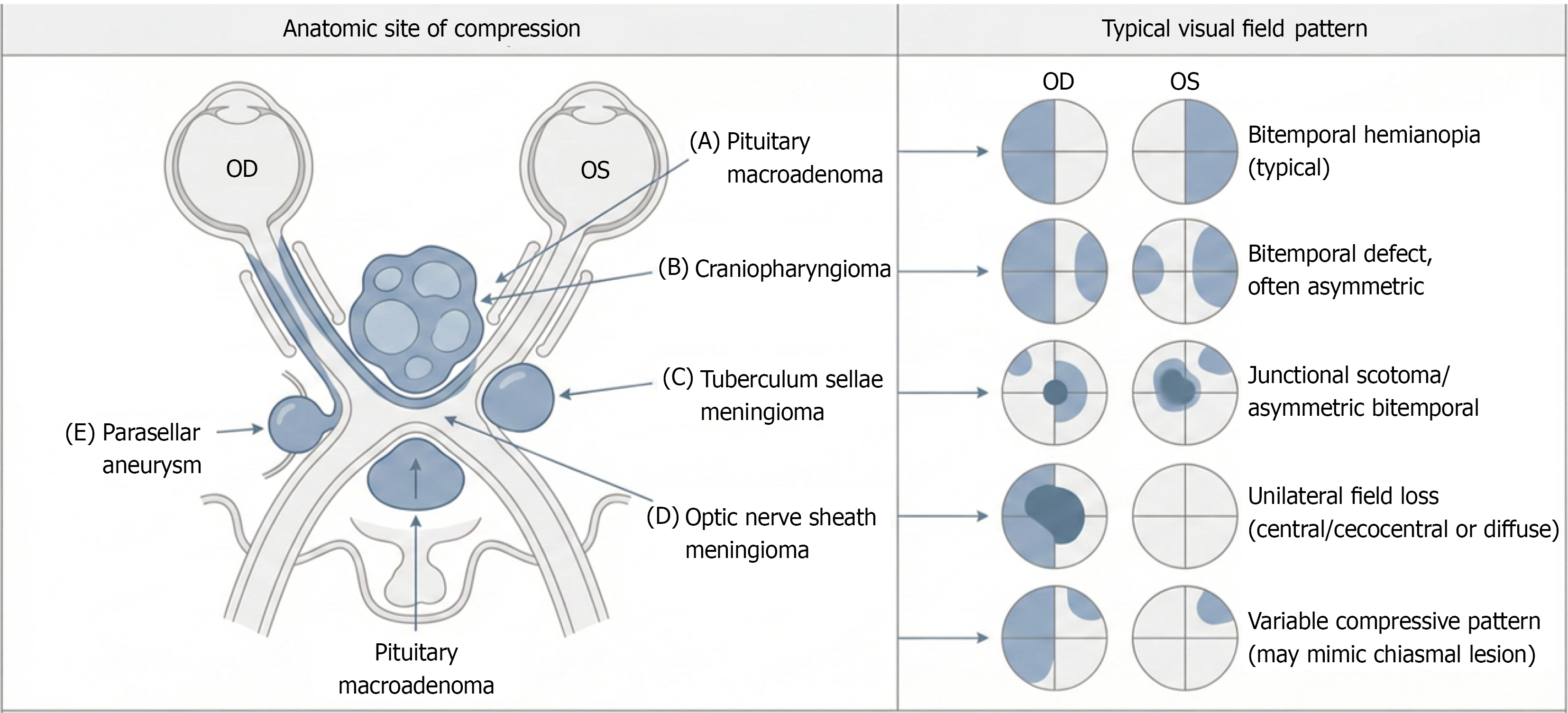
Figure 4 Compressive lesions along the optic nerve-chiasm axis and associated visual field patterns.
A: Pituitary macroadenoma; B: Craniopharyngioma; C: Tuberculum sellae meningioma; D: Optic nerve sheath meningioma; E: Parasellar aneurysm. OD: Oculus dexter; OS: Oculus sinister.
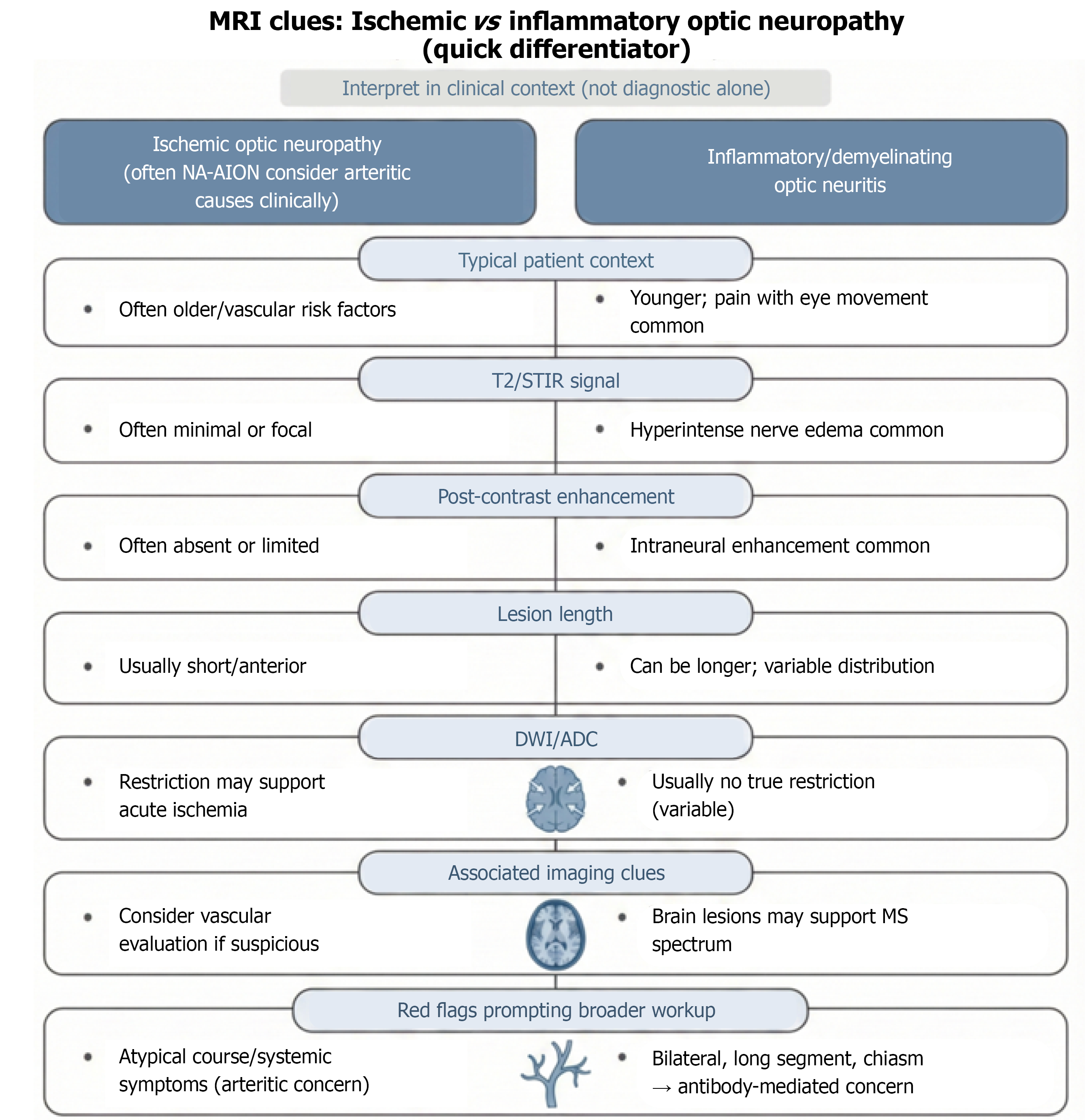
Figure 5 Magnetic resonance imaging features distinguishing ischemic optic neuropathy from inflammatory optic neuritis.
ADC: Apparent diffusion coefficient; DWI: Diffusion-weighted imaging; MRI: Magnetic resonance imaging; MS: Multiple sclerosis; NA-AION: Nonarteritic-anterior ischemic optic neuropathy; STIR: Short TI inversion recovery.
- Citation: Tlaiss Y, Hmayed J, Mneimneh M, Sarraf G, Gharios R, Zalzali I, Tarchichi A. Magnetic resonance imaging in the diagnosis of optic nerve and chiasmal pathologies. World J Ophthalmol 2026; 10(1): 119850
- URL: https://www.wjgnet.com/2218-6239/full/v10/i1/119850.htm
- DOI: https://dx.doi.org/10.5318/wjo.119850