
















Copyright: ©Author(s) 2026.
World J Ophthalmol. Sep 12, 2026; 10(1): 119850
Published online Sep 12, 2026. doi: 10.5318/wjo.119850
Published online Sep 12, 2026. doi: 10.5318/wjo.119850
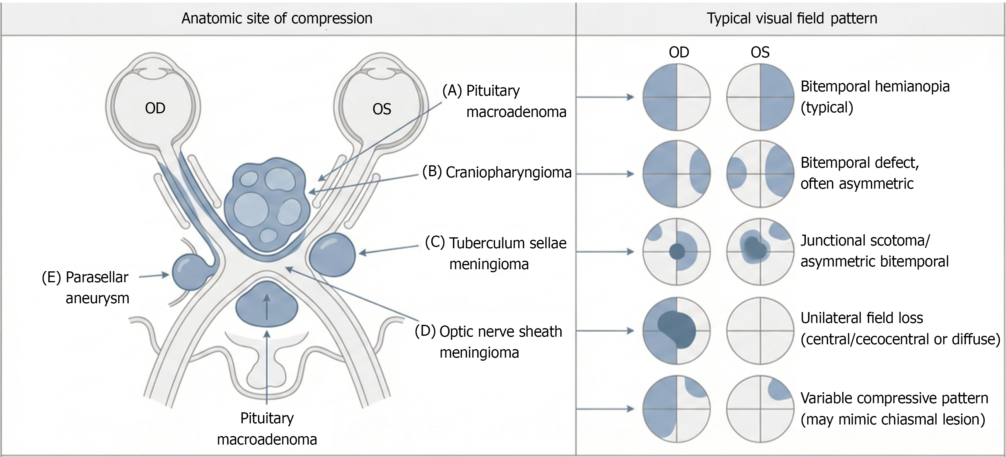
Figure 4 Compressive lesions along the optic nerve-chiasm axis and associated visual field patterns.
A: Pituitary macroadenoma; B: Craniopharyngioma; C: Tuberculum sellae meningioma; D: Optic nerve sheath meningioma; E: Parasellar aneurysm. OD: Oculus dexter; OS: Oculus sinister.
- Citation: Tlaiss Y, Hmayed J, Mneimneh M, Sarraf G, Gharios R, Zalzali I, Tarchichi A. Magnetic resonance imaging in the diagnosis of optic nerve and chiasmal pathologies. World J Ophthalmol 2026; 10(1): 119850
- URL: https://www.wjgnet.com/2218-6239/full/v10/i1/119850.htm
- DOI: https://dx.doi.org/10.5318/wjo.119850