Copyright
©2013 Baishideng Publishing Group Co.
World J Neurol. Dec 28, 2013; 3(4): 138-143
Published online Dec 28, 2013. doi: 10.5316/wjn.v3.i4.138
Published online Dec 28, 2013. doi: 10.5316/wjn.v3.i4.138
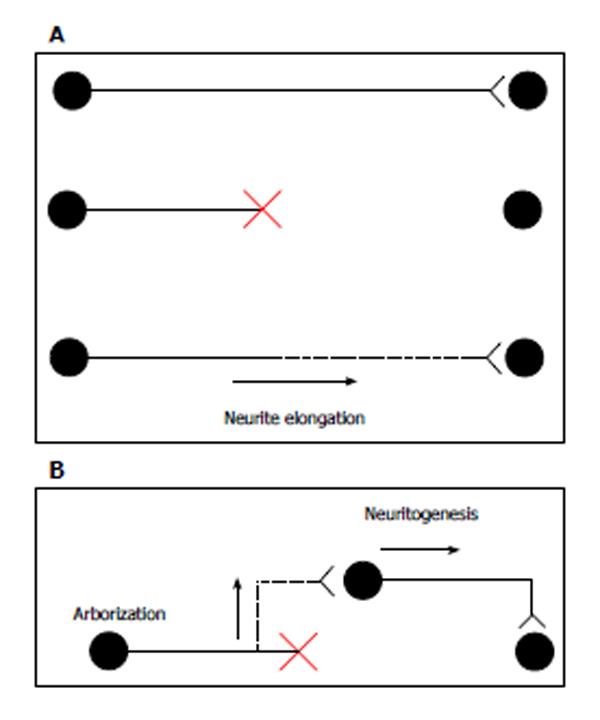
Figure 1 Schematic illustration of axonal regeneration.
A: Top, intact axon; middle, transected or crushed axon; bottom, canonical axon regeneration. New growth occurs from the tip of the transected axon, and the regenerating axon reinnervates its normal target; B: Regenerating axon: the branch arises from the axon close to the injury site. The new axon branch connects to the surrounding neuron, which extends an axon to the original target. Figures are modified from Tuszynski et al[50].
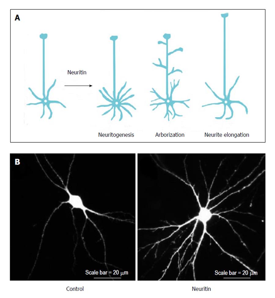
Figure 2 Neuritin induces neurite arborization.
A: Neuritin regulates neuritogenesis, neurite arborization, and neurite extension; B: Hippocampal neurons were prepared from rat embryos that were transfected with green fluorescent protein (GFP) (left) or GFP and neuritin (right) after 5 d in vitro, and then they were immunostained after 12 d in vitro.
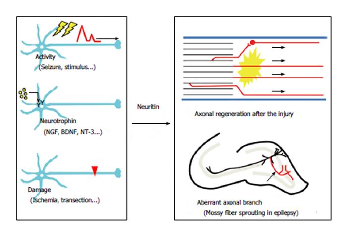
Figure 3 Induction of neuritin and its pathophysiological functions.
Representative neuronal events that increase the expression of neuritin. Seizures, neurotrophins, and neuronal damage induce the expression of neuritin (left). The physiological roles of neuritin. Neuritin-induced changes might promote axonal regeneration after nerve injury, whereas the same morphological changes could exacerbate temporal lobe epilepsy (right). NGF: Nerve growth factor; BDNF: Brain-derived neurotrophic factor; NT-3: Neurotrophin-3.
- Citation: Shimada T, Sugiura H, Yamagata K. Neuritin: A therapeutic candidate for promoting axonal regeneration. World J Neurol 2013; 3(4): 138-143
- URL: https://www.wjgnet.com/2218-6212/full/v3/i4/138.htm
- DOI: https://dx.doi.org/10.5316/wjn.v3.i4.138


