Copyright
©2013 Baishideng Publishing Group Co.
World J Neurol. Dec 28, 2013; 3(4): 138-143
Published online Dec 28, 2013. doi: 10.5316/wjn.v3.i4.138
Published online Dec 28, 2013. doi: 10.5316/wjn.v3.i4.138
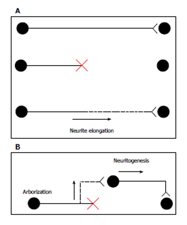
Figure 1 Schematic illustration of axonal regeneration.
A: Top, intact axon; middle, transected or crushed axon; bottom, canonical axon regeneration. New growth occurs from the tip of the transected axon, and the regenerating axon reinnervates its normal target; B: Regenerating axon: the branch arises from the axon close to the injury site. The new axon branch connects to the surrounding neuron, which extends an axon to the original target. Figures are modified from Tuszynski et al[50].
- Citation: Shimada T, Sugiura H, Yamagata K. Neuritin: A therapeutic candidate for promoting axonal regeneration. World J Neurol 2013; 3(4): 138-143
- URL: https://www.wjgnet.com/2218-6212/full/v3/i4/138.htm
- DOI: https://dx.doi.org/10.5316/wjn.v3.i4.138


