Copyright
©2014 Baishideng Publishing Group Inc.
World J Hematol. Nov 6, 2014; 3(4): 128-137
Published online Nov 6, 2014. doi: 10.5315/wjh.v3.i4.128
Published online Nov 6, 2014. doi: 10.5315/wjh.v3.i4.128
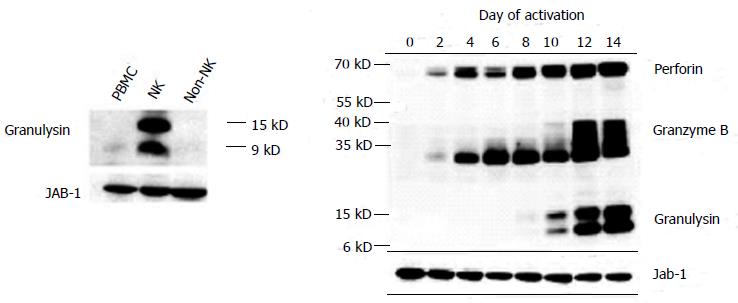
Figure 1 Expression of granulysin after T cell activation analyzed by western blotting.
Granulysin is expressed later compared to perforin and granzyme B after T cell activation. In natural killer (NK) cells, granulysin is constitutively expressed. Jab-1 (c-Jun activation domain-binding protein-1) is used as internal control. PBMC: Peripheral blood mononuclear cell.
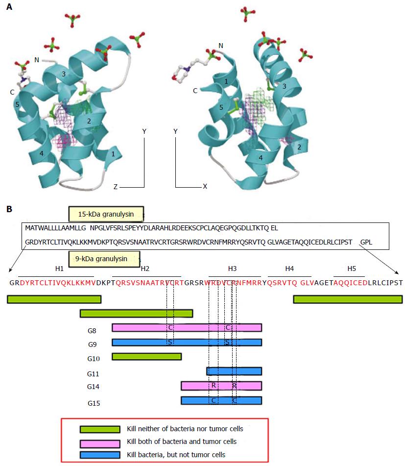
Figure 2 Granulysin.
A: 3-D structure model of 9-kDa granulysin. Granulysin consists of five a-helices. Cytotoxic active site ranges between helix-2 and helix-3, in which positive electric charges are located[9]; B: Scheme of cytotoxic active site in granulysin. Amino acid sequence of granulysin and its biologically active site are illustrated. See STRUCTURE AND FUNCTION in the text for detailed explanation.
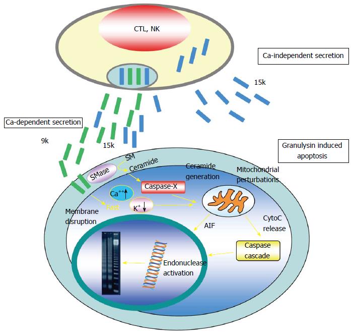
Figure 3 Schematic model of how granulysin kills target cell.
(Cited from ref.[48] revised by author). NK: Natural killer; AIF: Apoptosis-inducing factor; CTL: Cytotoxic T lymphocytes.
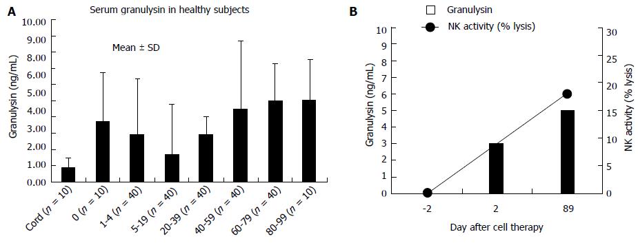
Figure 4 Serum granulysin in healthy subjects (A, see GRANULYSIN AS A BIOMARKER in the text) and relationship with natural killer cell activity (B).
Serum granulysin is increased along with the recovery of natural killer (NK) cell activity in infant with combined immunodeficiency after cell therapy.
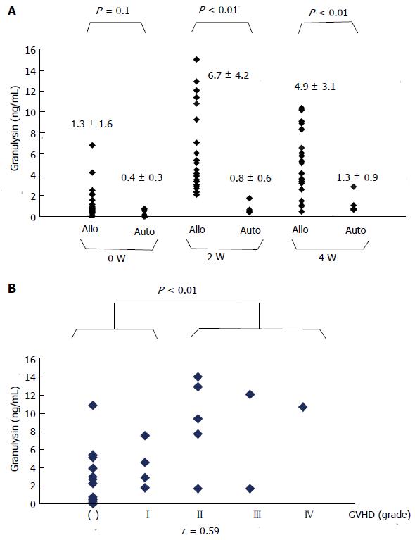
Figure 5 Trend of serum granulysin.
A: In patients with allo-hematopoietic stem cell transplantation (HSCT) (n = 21) and auto-HSCT (n = 5). Serum granulysin is elevated 2 wk after allogeneic hematopoietic stem cell transplantation, but not autologous hematopoietic stem cell transplantation; B: With the grade of graft-versus-host disease (GVHD). Serum granulysin and the grade of GVHD were plotted and their correlation coefficient was calculated (r = 0.59). The serum granulysin level of patients with grade 2 or more is significantly higher than that of patients with grade 1 or no GVHD.
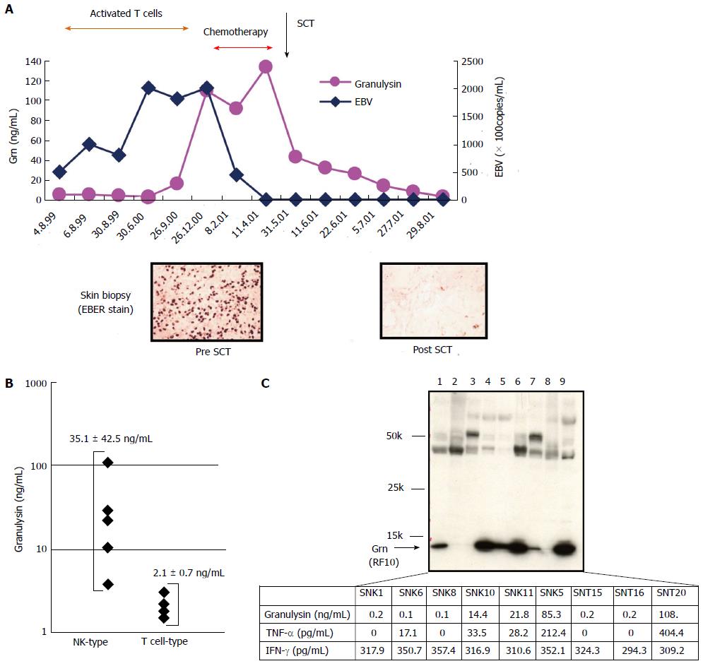
Figure 6 Serum granulysin.
A: Clinical course and trend of serum granulysin in a natural killer (NK) cell type chronic active EB virus infection (CAEBV) patient. For detailed explanation, see GRANULYSIN AS A BIOMARKER, 6: Granulysin in NK cell-related tumors or neoplasm in the text; B: Serum granulysin in patients with NK type and T cell type CAEBV, serum granulysin in patients with NK type (n = 5) and T cell type (n = 4) chronic active EB virus infection. Only in NK type CAEBV, serum granulysin is significantly elevated. Serum granulysin in patients with NK type (n = 5) and T cell type (n = 4) chronic active EB virus infection. Only in NK type CAEBV, serum granulysin is significantly elevated. C: Expression of granulysin and cytokine production in EB virus infected cell lines (SNK1,5,6,10,11: NK cell type, SNT8,15,16,20: γδT cell type). Western blotting was performed by using a monoclonal antibody, RF10 which reacts with 15-kDa but not 9-kDa granulysin. TNF-a and IFN-g in the culture supernatant were assayed by ELISA method. TNE: Tumor necrosis factor; INF: Interferon.
- Citation: Nagasawa M, Ogawa K, Nagata K, Shimizu N. Granulysin and its clinical significance as a biomarker of immune response and NK cell related neoplasms. World J Hematol 2014; 3(4): 128-137
- URL: https://www.wjgnet.com/2218-6204/full/v3/i4/128.htm
- DOI: https://dx.doi.org/10.5315/wjh.v3.i4.128


