
















©2014 Baishideng Publishing Group Inc.
World J Hematol. Nov 6, 2014; 3(4): 128-137
Published online Nov 6, 2014. doi: 10.5315/wjh.v3.i4.128
Published online Nov 6, 2014. doi: 10.5315/wjh.v3.i4.128
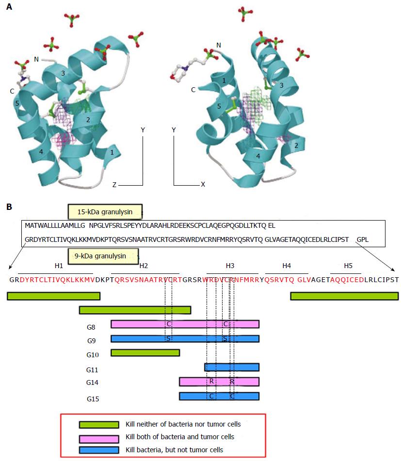
Figure 2 Granulysin.
A: 3-D structure model of 9-kDa granulysin. Granulysin consists of five a-helices. Cytotoxic active site ranges between helix-2 and helix-3, in which positive electric charges are located[9]; B: Scheme of cytotoxic active site in granulysin. Amino acid sequence of granulysin and its biologically active site are illustrated. See STRUCTURE AND FUNCTION in the text for detailed explanation.
- Citation: Nagasawa M, Ogawa K, Nagata K, Shimizu N. Granulysin and its clinical significance as a biomarker of immune response and NK cell related neoplasms. World J Hematol 2014; 3(4): 128-137
- URL: https://www.wjgnet.com/2218-6204/full/v3/i4/128.htm
- DOI: https://dx.doi.org/10.5315/wjh.v3.i4.128