Published online Nov 2, 2015. doi: 10.5314/wjd.v4.i4.145
Peer-review started: April 5, 2015
First decision: April 28, 2015
Revised: September 1, 2015
Accepted: October 16, 2015
Article in press: October 19, 2015
Published online: November 2, 2015
Processing time: 215 Days and 7.3 Hours
Cutaneous leiomyomas are rare, benign smooth muscle tumors, characterized by painful nodules in most of the cases. They can occur in multiple disseminated, segmental or zosteriform and solitary forms. Segmental or zosteriform leiomyoma can occur either alone (Type I), or with scattered nonsegmental lesions elsewhere (Type II); the latter variety occurring rarely. Here we present a case of Type II zosteriform leiomyoma in a middle aged individual.
Core tip: The most interesting feature of this case is the rarity of the presentation of the entity. Leiomyoma is not uncommon but zosteriform presentation is a relatively rare condition. Besides, discrete lesions apart from the zosteriform pattern, gives it the nomenclature of Type II zosteriform leiomyoma which is worth reporting.
- Citation: Das A, Podder I, Ghosh A. Zosteriform cutaneous leiomyoma-Type II: An uncommon presentation. World J Dermatol 2015; 4(4): 145-147
- URL: https://www.wjgnet.com/2218-6190/full/v4/i4/145.htm
- DOI: https://dx.doi.org/10.5314/wjd.v4.i4.145
Cutaneous leiomyomas are rare, benign painful tumors arising from smooth muscle cells. According to their site of origin, they are of three types; piloleiomyoma (most common variant), angioleiomyoma and genital leiomyoma[1]. These tumors can occur either singly or multiply. When multiple they can be arranged in diffuse (disseminated), blaschkoid or segmental (zosteriform) patterns[2]. Zosteriform leiomyomas commonly occur only along single dermatomes unilaterally (Type I); or in rare cases they may be associated with scattered, isolated, nonsegmental lesions elsewhere (Type II)[1]. Multiple leiomyomas may be associated with uterine leiomyomas and/or aggressive renal cell carcinomas. Here we present a case of Type II zosteriform leiomyoma; as this variety is extremely rare in occurrence.
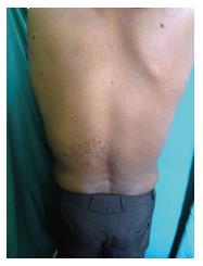
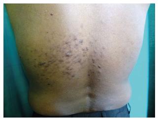
A 33-year-old male patient presented to us with multiple painful swellings, mainly on his back for the last 12 years. There were a few isolated lesions on the upper portion of his back, while several lesions were grouped together, near the middle portion, only on the left side (Figure 1). The isolated, scattered lesions developed earlier; all the lesions progressively increased in size over time. The patient also gave history of associated paroxysmal burning pain, which exacerbated on exposure to cold, touch or physical stress. Worsening of pain with cold was elicited. There was no history of any urinary disturbance. Family history was unremarkable. Cutaneous examination, revealed the lesions to be skin colored to erythematous, firm, tender papules and nodules, varying in size from 0.5 to 2 cm. The grouped lesions were present along the T9 and T10 dermatomes, in a segmental or zosteriform distribution, on the posterior aspect, unilaterally only over the left side (Figure 2). Few scattered isolated lesions were present on the upper part of back, shoulder distributed bilaterally. Hair, nails and mucosae were normal. Systemic examination was non contributory. Routine blood biochemical investigations did not reveal any abnormality. Ultrasound examination of the lower abdomen showing the kidney, ureter, bladder region was within normal limits. Human immunodeficiency virus serology was non reactive. Two nodules (zosteriform distribution and isolated) were subjected to excisional biopsy and histopathological examination (HPE) which showed bundles of spindle-shaped cells arranged in an interlacing and whorled pattern with elongated nuclei having rounded ends confirming the diagnosis of piloleiomyoma (Figure 3). The patient was started on nifedipine 10 mg thrice-daily, and advised periodic follow-up. The pain has decreased to a significant extent.
Cutaneous leiomyomas are benign smooth muscle tumors, which may occur singly or multiply. According to their site of origin they can be classified into three types[1,2]: (1) Piloleiomyoma - derived from the arrector pili muscles of hair follicles; (2) Angioleiomyoma (vascular leiomyoma) - arising from the tunica media of veins; and (3) Genital leiomyoma - mainly arising from the dartos muscle of scrotum; rarely they may be present over the labia majora and nipples[1]. Piloleiomyoma is the most common type, frequently occurring multiply.
Multiple cutaneous leiomyoma is the most common clinical variety, most commonly situated over the trunk and extremities. However, rarely they may occur on the tongue or any other part of mouth[1]. These lesions can be arranged in diffuse (disseminated), blaschkoid, linear or segmental (zosteriform) patterns[3]. Segmental or zosteriform leiomyoma can be classified into two types based on pathogenesis as proposed by Happle[4]; in Type I segmental leiomyoma there is a novel post zygotic segmental mutation; clinically characterized by the presence of dermatomal distribution only, while in Type II segmental leiomyoma there is post zygotic mutation in a heterogeneous embryo with subsequent loss of heterozygosity, resulting in segmental lesions along scattered, isolated, nonsegmental lesions. The index case had both discrete lesions as well as in a segmental or zosteriform pattern, unilaterally over the left side; thus a diagnosis of Type II segmental pilar leiomyoma was attained. Type II segmental leiomyoma has been rarely reported in the literature, which is commonly unilateral along a single dermatome[4,5]. However, bilateral involvement[6] and unilateral multisegmental distribution[2] have recently being reported. A loss of function mutation in the gene encoding fumarate hydratase on chromosome 1q has been found to predispose individuals to Type II segmental leiomyoma[7].
The lesions of leiomyoma are frequently painful, which may occur due to local pressure exerted by the tumor on cutaneous nerves or due to degranulation of infiltrating mast cells, or due to muscle contraction; the exact cause is yet to be elucidated. Multiple piloleiomyomas may be associated with several conditions viz. Reed’s syndrome (multiple cutaneous and uterine leiomyomas, hereditary leiomyomatosis and renal cell carcinoma)[1], erythrocytosis/polycythemia, and visceral involvement (gastrointestinal tract and retroperitoneal area)[4], etc.. Our patient has been advised regular follow up to detect any sign of RCC at the earliest. As leiomyoma is a painful tumor, all other painful cutaneous tumours may be considered as differential diagnoses; as summed up by the mnemonic: “blend an egg” representing: Blue rubber bleb naevus, leiomyoma, eccrine spiradenoma, neuroma, dermatofibroma, angiolipoma, neurilemmoma, endometrioma, glomus tumor and granular cell tumor[8]. Apart from the characteristic clinical features, HPE also clinched our diagnosis in favour of leiomyoma.
Treatment of the leiomyomas is not satisfactory. For solitary lesions curative surgery is the treatment of choice, preferably with preoperative embolization to minimize perioperative blood loss; whereas in case of extensive lesions various agents have been used to alleviate the associated pain like nifedipine, doxazosin, gabapentine, topical 9% hyoscine hydrobromide[2], phenoxybenzamine and botulinum toxin[9]. Liquid-nitrogen cryotherapy and CO2 laser ablation have been used with varying success[4]. Besides, there are reports of cases successfully treated with oral doxazosin[10]. Our patient was started on oral nifedipine, and advised regular follow-up.
Multiple painful swellings on the back for the preceding 12 years.
Leiomyoma.
Leiomyoma, neurofibroma, and eccrine spiradenoma.
Routine blood biochemical investigations did not reveal any abnormality. Human immunodeficiency virus serology was non-reactive.
Ultrasound examination of the lower abdomen showing the kidney, ureter, bladder region was within normal limits.
Histopathological examination which showed bundles of spindle-shaped cells arranged in an interlacing and whorled pattern with elongated nuclei having rounded ends confirming the diagnosis of piloleiomyoma.
Patient was prescribed oral nifedipine, and advised regular follow-up.
Sahoo B reported a case of Zosteriform pilar leiomyoma. A case of unilateral segmental Type II leiomyomatosis Vasani RJ.
A clinical differential diagnosis of leiomyoma should be considered in a patient presenting with zosteriform distribution of painful papules and nodules.
It is an interesting case presented, as long as there are only a few reported with zosteriform distribution.
P- Reviewer: Firooz A, González-López MA, Kaliyadan F, Manolache L S- Editor: Ji FF L- Editor: A E- Editor: Lu YJ
| 1. | James WD, Elston DM, Berger TG. Andrews’ Diseases of the skin: Clinical Dermatology. 11th ed. UK: Saunders Elsevier 2011; 614-615. |
| 2. | Kudligi C, Khaitan BK, Bhagwat PV, Asati DP. Unilateral multi-segmental leiomyomas: a report of rare case. Indian J Dermatol. 2013;58:160. [RCA] [PubMed] [DOI] [Full Text] [Cited by in Crossref: 1] [Cited by in RCA: 2] [Article Influence: 0.2] [Reference Citation Analysis (0)] |
| 3. | Sahoo B, Radotra BD, Kaur I, Kumar B. Zosteriform pilar leiomyoma. J Dermatol. 2001;28:759-761. [PubMed] |
| 4. | Gupta R, Singal A, Pandhi D. Skin-colored nodules in zosteriform pattern. Indian J Dermatol Venereol Leprol. 2005;72:81-82. [PubMed] |
| 5. | Vasani RJ. Unilateral segmental type II leiomyomatosis: a rare occurrence. Indian J Dermatol Venereol Leprol. 2012;78:521. [RCA] [PubMed] [DOI] [Full Text] [Cited by in RCA: 1] [Reference Citation Analysis (0)] |
| 6. | Suwattee P, Dakin C. Bilateral segmental leiomyomas: a case report and review of the literature. Cutis. 2008;82:33-36. [PubMed] |
| 7. | Parmentier L, Tomlinson I, Happle R, Borradori L. Evidence for a new fumarate hydratase gene mutation in a unilateral type 2 segmental leiomyomatosis. Dermatology. 2010;221:149-153. [RCA] [PubMed] [DOI] [Full Text] [Cited by in Crossref: 3] [Cited by in RCA: 4] [Article Influence: 0.3] [Reference Citation Analysis (0)] |
| 8. | DebBarman P, Das A, Shome K, Gharami RC. SkIndia Quiz 14: Solitary painful skin-colored nodule over the chest. Indian Dermatol Online J. 2014;5:354-355. [RCA] [PubMed] [DOI] [Full Text] [Full Text (PDF)] [Cited by in RCA: 1] [Reference Citation Analysis (0)] |
| 9. | Onder M, Adişen E. A new indication of botulinum toxin: leiomyoma-related pain. J Am Acad Dermatol. 2009;60:325-328. [RCA] [PubMed] [DOI] [Full Text] [Cited by in Crossref: 24] [Cited by in RCA: 27] [Article Influence: 1.7] [Reference Citation Analysis (0)] |
| 10. | Chaves AJ, Fernández-Recio JM, de Argila D, Rodríguez-Nevado I, Catalina M. [Zosteriform cutaneous leiomyoma. Satisfactory treatment with oral doxazosin]. Actas Dermosifiliogr. 2007;98:494-496. [PubMed] |


