Copyright
©2014 Baishideng Publishing Group Inc.
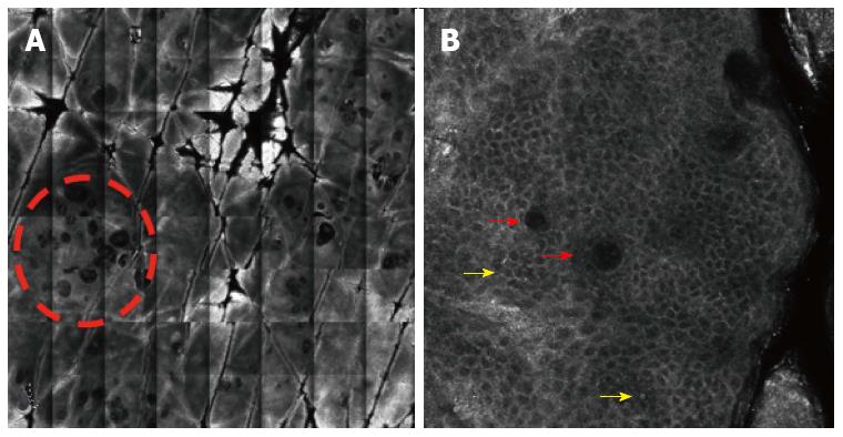
Figure 2 Image.
A: Vivablock image (4 mm × 4 mm) shows the spinous layer in a process of allergic contact dermatitis. Note the presence of multiple microvesicles (dashed circle); B: Reflectance confocal microscopy image (0.5 mm × 0.5 mm) reveals spongiosis and exocytosis (yellow arrow); in addition, microvesicle with lymphocytes inside (red arrow).
- Citation: Suárez-Pérez JA, Bosch R, González S, González E. Pathogenesis and diagnosis of contact dermatitis: Applications of reflectance confocal microscopy. World J Dermatol 2014; 3(3): 45-49
- URL: https://www.wjgnet.com/2218-6190/full/v3/i3/45.htm
- DOI: https://dx.doi.org/10.5314/wjd.v3.i3.45


