Copyright
©2014 Baishideng Publishing Group Inc.
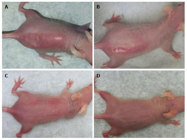
Figure 1 d-Limonene prevented sunburn formation.
Mice were exposed orally to 20 μL of pure or diluted d-limonene daily for 4 consecutive days followed by 1.5 kJ/ m2 of ultraviolet B exposure generated by a commercial sunlamp 1 d later. The maximum sunburn response occurred as shown by skin erythema on 6th day of UVR exposure was completely eliminated by pure d-limonene. A: UVR alone; B: UVR + 1.0% d-limonene in liponate; C: UVR + 10% d-limonene in liponate; D: UVR + 100% d-limonene. Topical application of d-limonene followed by UVR yielded similar results (data not shown). UVR: Ultraviolet irradiation.
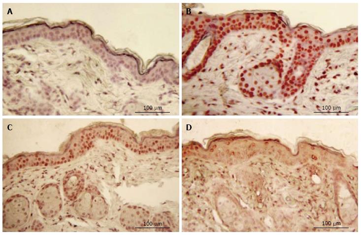
Figure 2 Immunostaining for cyclobutane pyrimidine dimers following oral exposure to d-limonene.
Skin samples were collected 5 min after UVR exposure. (A) Only unstained, CPD-negative cells are seen in control epidermis (B) intense CPD-stained nuclei are seen in skin samples (C, D). Oral d-limonene prevented CPD photoproducts formation in dose-dependent manner. UVR: Ultraviolet irradiation; CPD: Cyclobutane pyrimidine dimer.
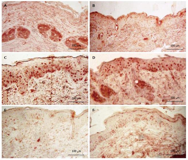
Figure 3 Immunostaining of N-myc downstream regulating gene 1.
Skin samples were obtained 6 d after UVR exposure. A: In control skin little or no NDRG1 was seen in epidermis but low levels were detected in sebaceous glands, Control; B: 100% d-limonene alone, mouse skin exposed to UVR strongly expressed NDRG1 both in epidermis and sebaceous glands (C-E); C: UVR alone; D: UVR + 1.0% d-limonene; E: UVR + 10% d-limonene; F: UVR + 100% d-limonene. Skin exposed to UVR with prior treatment of d-limonene, show significantly reduced NDRG1protein in epidermis and hair follicles (F). UVR: Ultraviolet irradiation; NDRG1: N-myc downstream regulating gene 1.
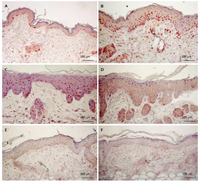
Figure 4 Immunostaining of proliferating cell nuclear antigen.
Skin samples were obtained on 6 d after UVR exposure. A: Control; B: 100% d-limonene alone; C: UVR alone; D: UVR + 1.0% d-limonene; E: UVR + 10% d-limonene; F: UVR + 100% d-limonene. (A) In control skin few PCNA-positive cells are observed in epidermis. Both 100% d-limonene alone (B) and UVR alone (C) induced proliferative stimulation in epidermis and hair follicles. However, pre-treatment of mice with d-limonene significantly reduced the PCNA-positive cells in skin compared to UVR-only mice (D-F), which verifies the CPD finding that oral d-limonene was protective against UVR-induced tissue damage. UVR: Ultraviolet irradiation; PCNA: Proliferating cell nuclear antigen; CPD: Cyclobutane pyrimidine dimer.
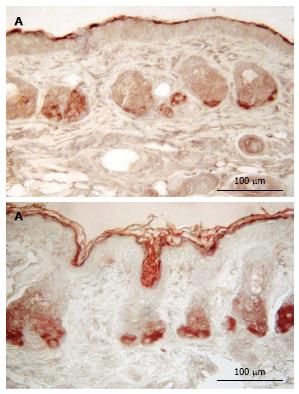
Figure 5 d-Limonene induced the expression of filaggrin.
A: Control; B: 100% d-limonene alone. Sebaceous glands of d-limonene–treated mice show a higher filaggrin signal compared to control mice. Also shown is the typical increased thickness of filaggrin-negative materials distal to the dark, thin filaggrin-positive layer, in comparison to control skin.
-
Citation: Uddin AN, Wu F, Labuda I, Tchou-Wong KM, Burns FJ.
d -limonene prevents ultraviolet irradiation: Induced cyclobutane pyrimidine dimers in Skh1 mouse skin. World J Dermatol 2014; 3(3): 64-72 - URL: https://www.wjgnet.com/2218-6190/full/v3/i3/64.htm
- DOI: https://dx.doi.org/10.5314/wjd.v3.i3.64


