Published online Feb 18, 2016. doi: 10.5312/wjo.v7.i2.117
Peer-review started: May 15, 2015
First decision: September 29, 2015
Revised: November 18, 2015
Accepted: December 3, 2015
Article in press: December 4, 2015
Published online: February 18, 2016
Processing time: 277 Days and 13.5 Hours
AIM: To evaluate the effect of different elbow and forearm positions on radiocapitellar alignment.
METHODS: Fifty-one healthy volunteers were recruited and bilateral elbow radiographs were taken to form a radiologic database. Lateral elbow radiographs were taken with the elbow in five different positions: Maximal extension and forearm in neutral, maximal flexion and forearm in neutral, elbow at 90° and forearm in neutral, elbow at 90° and forearm in supination and elbow at 90° and forearm in pronation. A goniometer was used to verify the accuracy of the elbow’s position for the radiographs at a 90° angle. The radiocapitellar ratio (RCR) measurements were then taken on the collected radiographs using the SliceOmatic software. An orthopedic resident performed the radiographic measurements on the 102 elbows, for a total of 510 lateral elbow radiographic measures. ANOVA paired t-tests and Pearson coefficients were used to assess the differences and correlations between the RCR in each position.
RESULTS: Mean RCR values were -2% ± 7% (maximal extension), -5% ± 9% (maximal flexion), and for elbow at 90° and forearm in neutral -2% ± 5%, supination 1% ± 6% and pronation 1% ± 5%. ANOVA analyses demonstrated significant differences between the RCR in different elbow and forearm positions. Paired t-tests confirmed significant differences between the RCR at maximal flexion and flexion at 90°, and maximal extension and flexion. The Pearson coefficient showed significant correlations between the RCR with the elbow at 90° - maximal flexion; the forearm in neutral-supination; the forearm in neutral-pronation.
CONCLUSION: Overall, 95% of the RCR values are included in the normal range (obtained at 90° of flexion) and a value outside this range, in any position, should raise suspicion for instability.
Core tip: Assessing radial head alignment after injury and obtaining perfect lateral radiographs with the elbow at 90° and the forearm in neutral may be difficult. Therefore we designed this study to assess whether the radiocapitellar ratios (RCR) calculated from true lateral radiographs at different positions of elbow flexion and forearm pronosupination differ from those taken in 90° flexion and neutral position. The paper shows that the RCR measurement continues to be an overall valid and reliable method throughout different elbow and forearm positions. However, values in the negative range, > 5% regardless of forearm rotation, should raise suspicion for elbow instability.
- Citation: Sandman E, Canet F, Petit Y, Laflamme GY, Athwal GS, Rouleau DM. Effect of elbow position on radiographic measurements of radio-capitellar alignment. World J Orthop 2016; 7(2): 117-122
- URL: https://www.wjgnet.com/2218-5836/full/v7/i2/117.htm
- DOI: https://dx.doi.org/10.5312/wjo.v7.i2.117
The elbow is a complex joint that is comprised of three articulations: The ulno-humeral, the radiocapitellar and the proximal radio-ulnar joints. The joint capsule and the ligamentous structures surrounding the elbow’s congruent osseous articulations provide static stability while its adjacent muscles and tendons offer dynamic stability by aligning and compressing the joint surfaces together[1]. The components of elbow stability can be divided into primary and secondary stabilizers. The elbow’s primary stabilizers consist of the anterior bundle of the medial collateral ligament, the lateral ulnar collateral ligament, and the ulnohumeral joint[2]. The secondary stabilizers involve the radial head, the joint capsule and the adjacent muscles surrounding the articulation. All of these structures function together to permit functional elbow flexion-extension and forearm pronation-supination ranges of motion (ROM). However, elbow stability and alignment can easily be disrupted after a trauma. In fact, the elbow is second only to the shoulder for the incidence of non-prosthetic joint dislocation[3].
The literature highlights the importance of evaluating a joint’s integrity throughout its full arc of movement, as the stability of an articulation is a dynamic process. Assessing an articulation with a single radiologic view may lead to suboptimal diagnostics and treatments. Therefore, the evaluation of the radiocapitellar joint, which is known to contribute to elbow stability, would be an added resource. In their study of 80 healthy elbows, Rouleau et al[4] described a quantitative method to assess radiocapitellar joint translations, the radiocapitellar ratio (RCR), defined as the displacement of the radial head (minimal distance between the right bisector of the radial head and the center of the capitellum) divided by the diameter of the capitellum[4]. The mean normal RCR was 4% ± 4% (95%CI: -5% to 13%). It has been reported to have good inter- and intra-observer reliability when measured on a lateral radiograph with the elbow positioned at 90° of flexion with neutral forearm rotation. In a trauma setting, it may be difficult to obtain standardized lateral radiographs with the elbow flexed at 90° and the forearm in neutral rotation due to factors such as pain, swelling, or fractures[1], which may cause radiographs to be taken with the elbow and the forearm in different positions. The purpose of this study was to assess whether RCRs calculated from true lateral radiographs, at different positions of elbow flexion and forearm pronosupination, differ from those taken in 90° flexion and neutral position.
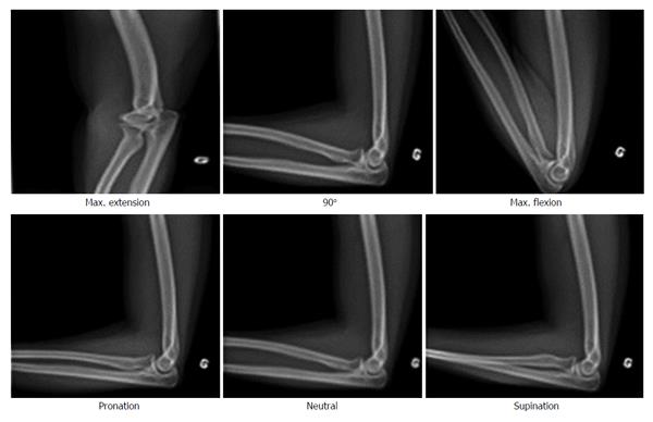
Fifty-one healthy volunteers were recruited and bilateral elbow radiographs were taken to form a radiologic database. In this study, the volunteers included 31 females and 20 males, with an average age of 32 years old (SD = 9.0). The number of radiographs observed followed the guidelines of Harrison et al[5]. The inclusion criteria were: patients aged between 18-50 years old, and the absence of a preexisting elbow pathology in both upper extremities. The exclusion criteria consisted of: Elbows with preexisting abnormalities, such as arthrosis, fractures, surgical implants, etc., and pregnant women or those at risk of being pregnant. Each individual was asked to give informed consent and protected with lead aprons. They were asked to actively move their elbow into the various positions, so that no passive maximal pressure was applied. Ninety degree elbow flexion was assured by measuring with a goniometer at the time of imaging and was reviewed during measurements on the computer software. Lateral elbow radiographs were taken with the elbow in five different positions: Maximal extension and forearm in neutral, maximal flexion and forearm in neutral, elbow at 90° and forearm in neutral, elbow at 90° and forearm in supination and elbow at 90° and forearm in pronation (Figure 1). As described by London et al[6] a true lateral elbow radiograph was achieved when the trochlear sulcus, the capitellum and the medial trochlea were concentrically superimposed. The Institutional Review Board of the ethical committee approved this study.
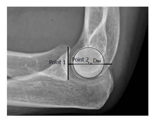
The RCR method was used to measure the translation of the radial head on the capitellum, described in 5 steps[4], with SliceOmatic (Tomovision Inc, Magog, Quebec, Canada) software: (1) A line, perpendicular to the joint, was drawn at the center of the articular surface of the radial head (Figure 2, point 1); (2) The diameter of the capitellum (Ø capitellum) was measured; (3) The center of the capitellum was identified as the bisector of the capitellum’s diameter (Figure 2); (4) The minimal distance between the center points of the radial head and the capitellum was measured (Figure 2); and (5) The Radial-Capitellum-Ratio was calculated: RCR (%) = DRH/Øcapitellum.
A positive RCR value indicates anterior radial head translation, while a negative RCR result signifies posterior radial head translation. An orthopedic resident (ES) performed the radiographic measurements on the 102 elbows, for a total of 510 lateral elbow radiographic measures. The intra-observer (0.72) and inter-observer reliability (0.52) of this method were previously reported using intraclass correlation tests[4]. The results obtained were compared to the normal RCR range, measured in the previous study by Rouleau et al[4] and described as a RCR value between -5% to 13%. In their study, the measurements were taken twice by two different observers and the mean normal RCR was 4% ± 4%, with the normal RCR range within a 95%CI.
ANOVA and paired t tests were used to assess the differences in RCR measurement results between the five different elbow and forearm positions, with a level of significance established at P < 0.05. Pearson coefficients were calculated to assess the correlation between the RCR measurements in each different elbow and forearm position. Correlation coefficients (r) were considered small if r = ± 0.00 to 0.09; medium if r = ± 0.10 to 0.30; and strong if r = ± 0.50 and 1.00[7]. According to the results of the mean and standard deviation, analyses of the power for the Pearson coefficients correlations were also calculated. Statistical review of the study was performed by a biomedical statistician.
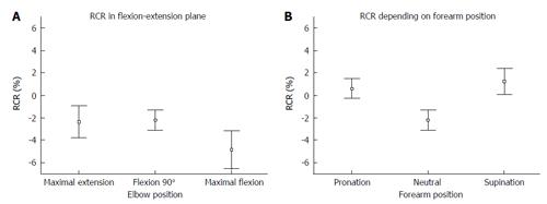
The mean maximal flexion achieved by the 51 subjects was of 151°± 5° and the mean maximal extension was of 12°± 7°. The mean RCRs for each position were: elbow in maximal extension: -2% ± 7% (95%CI: -4% to -1%), elbow in maximal flexion: -5% ± 9% (95%CI: -6% to -3%), elbow at 90° and forearm in neutral: -2% ± 5% (95%CI: -3% to -1%), elbow at 90° and forearm in supination: 1% ± 6% 95%CI: 0% to 2%), and elbow at 90° and forearm in pronation: 1% ± 5% (95%CI: 0% to 2%) (Figure 3). According to the ANOVA results, a significant difference exists between the RCRs in different elbow positions (P = 0.01) and in different forearm positions (P < 0.001). Moreover, 95% of our cohort obtained RCR values between the normal ranges initially evaluated, with posterior translation of the radial head of 5% to anterior translation of 13%.
Paired t tests were used to accommodate the fact that these are non-independent events, and confirmed a significant difference between maximal elbow flexion and 90° of elbow flexion (P = 0.003), as well as for maximal elbow extension and maximal elbow flexion (P = 0.034) (Table 1). Additionally, the paired t test showed significant differences between the positions of the forearm in neutral and pronation (P≤ 0.001), as well as between the forearm in neutral and supination (P < 0.001). However, there was no significant difference between the positions of elbow flexion at 90° and maximal extension (P = 0.86), nor between the positions of the forearm in pronation and in supination (P = 0.28).
| Paired t-test for elbow and forearm positions | P |
| Maximal elbow extension and maximal elbow flexion | 0.034 |
| Maximal elbow flexion and elbow flexion at 90° | 0.003 |
| Maximal elbow extension and elbow flexion at 90° | 0.86 |
| Forearm in neutral and forearm in pronation | 0.001 |
| Forearm in neutral and forearm in supination | 0.001 |
| Forearm in pronation and forearm in supination | 0.28 |
According to the Pearson coefficients, significant correlations exist between elbow flexion at 90° and in maximal flexion (r = 0.19, P = 0.049), the forearm in neutral and in supination (r = 0.34, P < 0.001), as well as the forearm in neutral and in pronation (r = 0.42, P < 0.001).
There was no significant correlation observed between the forearm positions in pronation and supination (r = 0.37, P = 0.55), the elbow positioned at 90° and in maximal extension (r = 0.086, P = 0.39) or between maximal elbow flexion and maximal elbow extension (r =0.085, P = 0.39).
Post hoc power analyses of the Pearson coefficient correlations were done for the different elbow and forearm positions (Table 2). Significant power was only obtained when comparing maximal elbow flexion and maximal elbow extension (Π = 0.84). The power calculated for elbow flexion at 90° with the forearm in neutral and maximal elbow extension was Π = 0.63, and Π = 0.05 for elbow flexion at 90° with the forearm in neutral and maximal elbow flexion. When analyzing the power for the different forearm positions, significant results were obtained when comparing pronation and neutral, as well as between supination and neutral forearm positions, both with a power Π = 0.99. The power found for the correlation between supination and pronation forearm positions was 0.18.
| r | P | Power (Π) | |
| Pearson coefficient correlation for the elbow | |||
| Maximal extension and Maximal flexion | -0.0854 | 0.394 | 0.84 |
| Maximal flexion and flexion at 90° | 0.1948 | 0.050 | 0.05 |
| Maximal extension and flexion at 90° | -0.0860 | 0.390 | 0.63 |
| Pearson coefficient correlation for the forearm | |||
| Neutral and pronation | 0.42 | 0.001 | 0.99 |
| Neutral and supination | 0.34 | 0.001 | 0.99 |
| Pronation and supination | 0.37 | 0.55 | 0.18 |
Following upper extremity trauma, a complete evaluation of the elbow’s primary and secondary stabilizers is necessary to avoid occult injuries and inappropriate treatments. The stability of an articulation can be determined clinically or with radiographic imaging. In the trauma setting, an elbow’s clinical stability and complete ROM evaluation may be difficult due to associated injuries and pain. Perfect lateral radiologic views at 90° of flexion may also be difficult to obtain due to multiple factors. Cheung et al[8] described the importance of obtaining proper alignment on a lateral radiograph with the forearm in neutral, with views of both the elbow and the wrist. Moreover, it has been suggested in the literature that stability of the radial head, especially after reduction, should be evaluated throughout its full ROM under radiological imaging which is what would make the RCR value of interest.
When analyzing the results obtained with the paired t-tests, significant differences were found for the RCR measurements between maximal elbow flexion and elbow flexion at 90°; between maximal elbow flexion and maximal elbow extension; between neutral and pronation forearm positions; as well as between neutral and supination forearm positions. Thus, elbow and forearm positioning seem to substantially influence radiocapitellar alignment, because our results tend to demonstrate significant differences for most of the positions evaluated. Although these differences are statistically significant, further research is needed to evaluate if they are clinically important, as a RCR of 5% represents a small translation of the radial head (1.25 mm for a capitellum of 25 mm of diameter).
The RCR measurement method has previously been shown to be valid and reliable when evaluating translations of the radiocapitellar articulation, with the elbow at 90° and the forearm in neutral[4]. This study evaluated the RCR method in five different elbow and forearm positions. The different elbow positions seem to have a greater effect on the RCR measurement results, when compared to the different forearm ranges of motion. Nonetheless, 95% of our cohort obtained RCR values between the normal ranges initially evaluated from -5% to 13%[4]. To illustrate, this range corresponds, in a capitellum with a diameter of 25 mm, to a radiocapitellar translation of 1.25 mm posterior to 3.25 mm anterior, for a total average of 5 mm displacement. Thus, the RCR measurement continues to be an overall valid and reliable method throughout different elbow and forearm positions.
The main limitations of this study are that the radiographs were all taken with the radiological beam perpendicular to the elbow joint, to obtain a perfect lateral view. Further studies should be done to evaluate the effect of the radiological beam angle on the measurement of radial head displacement, since radiographs taken with mild misalignment or with the elbow slightly oblique might influence the measurements. Finally, an injured elbow may not be able to achieve the different elbow positions tested in the study, due to pain, swelling or altered mechanics. However, the positions were chosen to cover the entire range of motion of the elbow, as well as to maximize the differences on the RCR measurements.
To conclude, even if positioning is not ideal, if a true lateral radiograph of the elbow is taken, the RCR should fall within the normal range of -5% to 13% when the radiocapitellar joint is intact. The RCR measurement method is dependent on elbow (flexion-extension) and forearm (pronation-supination) positions. In both maximal elbow positions in flexion and extension, the measurements of the RCR have a higher standard deviation. In order to decrease its variability, we recommend, as a convention, measuring the RCR on lateral radiographs with the elbow at 90° and the forearm in any position (pronation, neutral or supination). In normal elbows, at 90° of flexion, the RCR measurement with the forearm in pronation and supination show a significant difference from the forearm in neutral, and move the RCR in a positive direction. Therefore values in the negative range, > 5% regardless of forearm rotation, should raise suspicion for instability. A clinical study on the prognosis value of RCR in the presence of acute elbow dislocation would further support its clinical utility[9].
The elbow is a complex joint that is comprised of three articulations and all of these structures function together to permit functional elbow flexion-extension and forearm pronation-supination ranges of motion (ROM). The literature highlights the importance of evaluating a joint’s integrity throughout its full arc of movement, since the stability of an articulation is a dynamic process. However, elbow stability and alignment can easily be disrupted after a trauma and few reliable measurement methods are available. The radiocapitellar ratio (RCR) was described as a quantitative method to assess radiocapitellar joint translation on standardized lateral radiographs with the elbow flexed at 90° and the forearm in neutral rotation. However, it may be difficult in a trauma setting to obtain perfect lateral radiographs. Thus, it was of interest to assess whether the RCRs calculated from true lateral radiographs, at different positions of elbow flexion and forearm pronation-supination, differ from those taken in 90° flexion and neutral position.
The authors aimed to evaluate the effect of different elbow and forearm positions on radiocapitellar alignment, using the RCR on fifty-one healthy volunteers. Bilateral elbow radiographs were taken with the elbow in five different positions to form a radiologic database to investigate if elbow position influenced the RCR.
This study demonstrate that even if positioning is not ideal, if a true lateral radiograph of the elbow is taken, the RCR should fall within the normal range of -5% to 13% when the radiocapitellar joint is intact. However, values in the negative range, > 5% regardless of forearm rotation, should raise suspicion for elbow instability or subluxation.
The authors believe that further studies should be done to evaluate the effect of the radiological beam angle on the measurement of radial head displacement, since radiographs taken with mild misalignment or with the elbow slightly oblique might influence the measurements.
A quantitative method to assess radiocapitellar joint translations, the RCR, is defined as the displacement of the radial head (minimal distance between the right bisector of the radial head and the center of the capitellum) divided by the diameter of the capitellum. The mean normal RCR is 4% ± 4% (95%CI: -5% to 13%). It has been reported to have good inter- and intra-observer reliability when measured on a lateral radiograph with the elbow positioned at 90 degrees of flexion with neutral forearm rotation.
The authors concur with the literature with regard to the importance of obtaining proper alignment on a lateral radiograph with the forearm in neutral, with views of both the elbow and the wrist. Moreover, stability of the radial head, especially after reduction, should be evaluated throughout its full ROM under radiological imaging which is what would make the RCR value of interest. Therefore, this review article may have potential to increase knowledge to optimize diagnosis and treatment of elbow injuries.
P- Reviewer: Malik H, Ranjan Patra S S- Editor: Qiu S L- Editor: A E- Editor: Liu SQ
| 1. | Herman MJ, Boardman MJ, Hoover JR, Chafetz RS. Relationship of the anterior humeral line to the capitellar ossific nucleus: variability with age. J Bone Joint Surg Am. 2009;91:2188-2193. [RCA] [PubMed] [DOI] [Full Text] [Cited by in Crossref: 53] [Cited by in RCA: 50] [Article Influence: 3.1] [Reference Citation Analysis (0)] |
| 2. | Athwal GS, Ramsey ML, Steinmann SP, Wolf JM. Fractures and dislocations of the elbow: a return to the basics. Instr Course Lect. 2011;60:199-214. [PubMed] |
| 3. | Coonrad RW, Roush TF, Major NM, Basamania CJ. The drop sign, a radiographic warning sign of elbow instability. J Shoulder Elbow Surg. 2005;14:312-317. [PubMed] |
| 4. | Rouleau DM, Sandman E, Canet F, Djahangiri A, Laflamme Y, Athwal GS, Petit Y. Radial head translation measurement in healthy individuals: the radiocapitellar ratio. J Shoulder Elbow Surg. 2012;21:574-579. [RCA] [PubMed] [DOI] [Full Text] [Cited by in Crossref: 11] [Cited by in RCA: 11] [Article Influence: 0.8] [Reference Citation Analysis (0)] |
| 5. | Harrison DE, Harrison DD, Cailliet R, Janik TJ, Holland B. Radiographic analysis of lumbar lordosis: centroid, Cobb, TRALL, and Harrison posterior tangent methods. Spine (Phila Pa 1976). 2001;26:E235-E242. [PubMed] |
| 6. | London JT. Kinematics of the elbow. J Bone Joint Surg Am. 1981;63:529-535. [PubMed] |
| 7. | Cohen J. Statistical Power Analysis for the Behavioral Sciences, 2nd edition. NY, New York: Routledge Academic 1988; . |
| 8. | Cheung EV, Yao J. Monteggia fracture-dislocation associated with proximal and distal radioulnar joint instability. A case report. J Bone Joint Surg Am. 2009;91:950-954. [RCA] [PubMed] [DOI] [Full Text] [Cited by in Crossref: 17] [Cited by in RCA: 19] [Article Influence: 1.2] [Reference Citation Analysis (0)] |
| 9. | Windisch G, Clement H, Grechenig W, Tesch NP, Pichler W. The anatomy of the proximal ulna. J Shoulder Elbow Surg. 2007;16:661-666. [PubMed] |


