Published online Nov 18, 2021. doi: 10.5312/wjo.v12.i11.954
Peer-review started: July 5, 2021
First decision: July 28, 2021
Revised: August 30, 2021
Accepted: September 27, 2021
Article in press: September 27, 2021
Published online: November 18, 2021
Processing time: 134 Days and 3.6 Hours
Monteggia fractures are uncommon injuries in paediatric age. Treatment algorithms assert that length-unstable fractures are treated with plate fixation. In this case report, intramedullary fixation of an acute length-unstable Monteggia fracture allowed a stable reduction to be achieved, along with an appropriate ulnar length and alignment as well as radio capitellar reduction despite the fact that the orthopaedic surgeon did not use a plate for the ulnar fracture. The scope of treatment is to avoid the use of a plate that causes periosteal stripping and blood circulation disruption around the fracture.
A four-year-old girl presented at the Emergency Department following an accidental fall off a chair onto the right forearm. The X-ray highlighted a length-unstable acute Bado type 1 Monteggia fracture of the right forearm. On the same day, the patient underwent surgical treatment of the Monteggia fracture. The surgeon preferred not to use a plate to avoid a delay in fracture healing and to allow the micromotion necessary for callus formation. The operation comprised percutaneous fixation with an elastic intramedullary K-wire of the ulnar fracture and, subsequently, humeroradial joint reduction through manual manipulation. The orthopaedic surgeon assessed the stability of the radial head reduction under fluoroscopic control through flexion, extension, pronation and supination of the forearm. Healing of the fracture occurred within six weeks after surgery, as indicated by the presence of calluses on at least three cortices on standard radiographs. Dislocation/subluxation or loss of ulnar reduction was not apparent at the final X-ray examination.
Intramedullary fixation of unstable Monteggia fractures results in excellent outcomes, provides reliable reduction and causes fewer complications.
Core Tip: Treatment algorithms assert that acute length-stable Monteggia fractures are treated with an intramedullary device, while acute length-unstable fractures are treated with plates. Intramedullary devices have the advantage of smaller skin incisions, less soft tissue disruption, shorter operative times and easier device removal. Plates allow more anatomical restoration of the ulnar fracture and the radial bow, although they can cause delayed union due to blood circulation disruption around the fracture. Intramedullary wires can be used for the treatment of acute length-unstable Monteggia fractures instead of plates as these are associated with excellent results and fewer complications.
- Citation: Evola FR, Di Fede GF, Bonanno S, Evola G, Cucuzza ME. Management of acute length-unstable Monteggia fractures in children: A case report . World J Orthop 2021; 12(11): 954-960
- URL: https://www.wjgnet.com/2218-5836/full/v12/i11/954.htm
- DOI: https://dx.doi.org/10.5312/wjo.v12.i11.954
Monteggia fractures are uncommon injuries in paediatric age. In the nineteenth century, an Italian surgeon named Giovanni Monteggia described a traumatic injury involving a fracture of the proximal ulna with an associated dislocation of the radial head and disruption of the radioulnar joint[1]. Subsequently, these fractures were classified by Bado[2] into four types according to the direction of dislocation of the radial head.
The mechanism of injury is caused by direct trauma and hyperpronation as well as hyperextension[3]. The occurrence of paediatric Monteggia fractures covers between 1. Of 5% and 3% of all childhood elbow injuries[3,4]. Monteggia fractures remain a challenge for paediatric orthopaedic surgeons because of the difficulty involved in diagnosis and in the treatment of missed radial head dislocation and late instability. The aim of treatment is to achieve stable reduction of the ulnar fracture and radial head dislocation. Nowadays, the surgical treatment modalities available for these acute fractures include plates, intramedullary Kirschnerwires or elastic stable intramedullary nailing and, more recently, external fixation devices[5]. Although plates and intramedullary devices have comparable outcomes and complications, nailing or K-wires have the advantage of smaller skin incisions, less soft tissue disruption, shorter operative times and easier device removal. Furthermore, intramedullary wire fixations, due to their lack of rigidity, allow micromotion and callus formation[5]. Mechanical properties of intramedullary wires are based on a three-point fixation of the inner cortex and on a spread of the interosseous membrane[5]. Nowadays, there is no consensus regarding the preferred nail diameter, although, in the literature, authors suggest that the diameter of the wire should be approximately two-thirds of the medullary canal, measured atthe isthmus level[6]. Intramedullary devices may cause skin irritation, refractures, malunion, secondary displacement and nerve injury. The use of plates is associated with a more anatomical restoration of the ulnar fracture and the radial bow, and a more rigid fixation, which requires reduced post-operative immobilization[6]. Despite this, open reduction and plate fixation cause periosteal stripping and blood circulation disruption around the fracture, resulting in a greater likelihood of delayed union or more rarely non-union[6].
The aim of this case report was to assess the efficacy of intramedullary wires for the treatment of acute length-unstable Monteggia fractures instead of plate fixation and to review the literature.
A four-year-old right-hand-dominant girl presented at the Emergency Department of our hospital following an accidental fall off a chair onto the right forearm. The mother reported a direct trauma to the floor of the right forearm with hyperextension and hyperpronation.
Post-trauma, the child reported pain in the right forearm along with inability to bend the elbow.
The patient had a free previous surgical history.
The child had forearm deformities with swelling and local tenderness around the elbow. Furthermore, active and passive motion of the elbow was impossible and accompanied by pain.
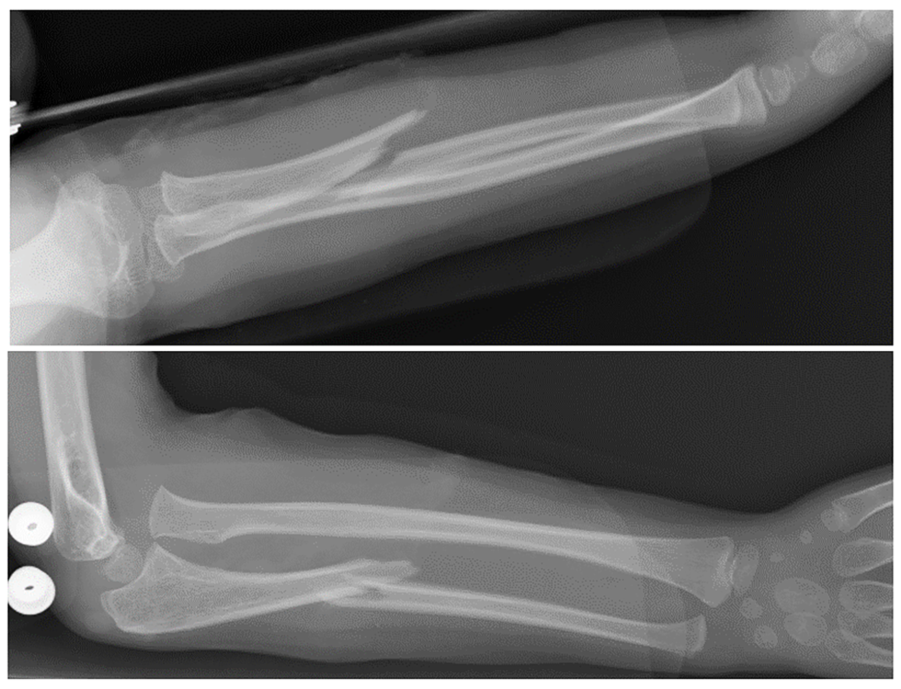
The X-rays appeared to show a fracture of the proximal ulna with an associated anterior dislocation of the radial head of the right elbow (Figure 1). The ulnar injury consisted of a long oblique fracture with a line measuring more than twice the cortical diameter. Radial head dislocation may be diagnosed when the radiocapitellar line, drawn through the axis of the radial neck on a lateral radiograph, regardless of the degree of flexion or extension of the elbow, crosses the humeral capitellum anterior to this normal position.
The X-ray highlighted a length-unstable acute Bado type1 Monteggia fracture of the right forearm.
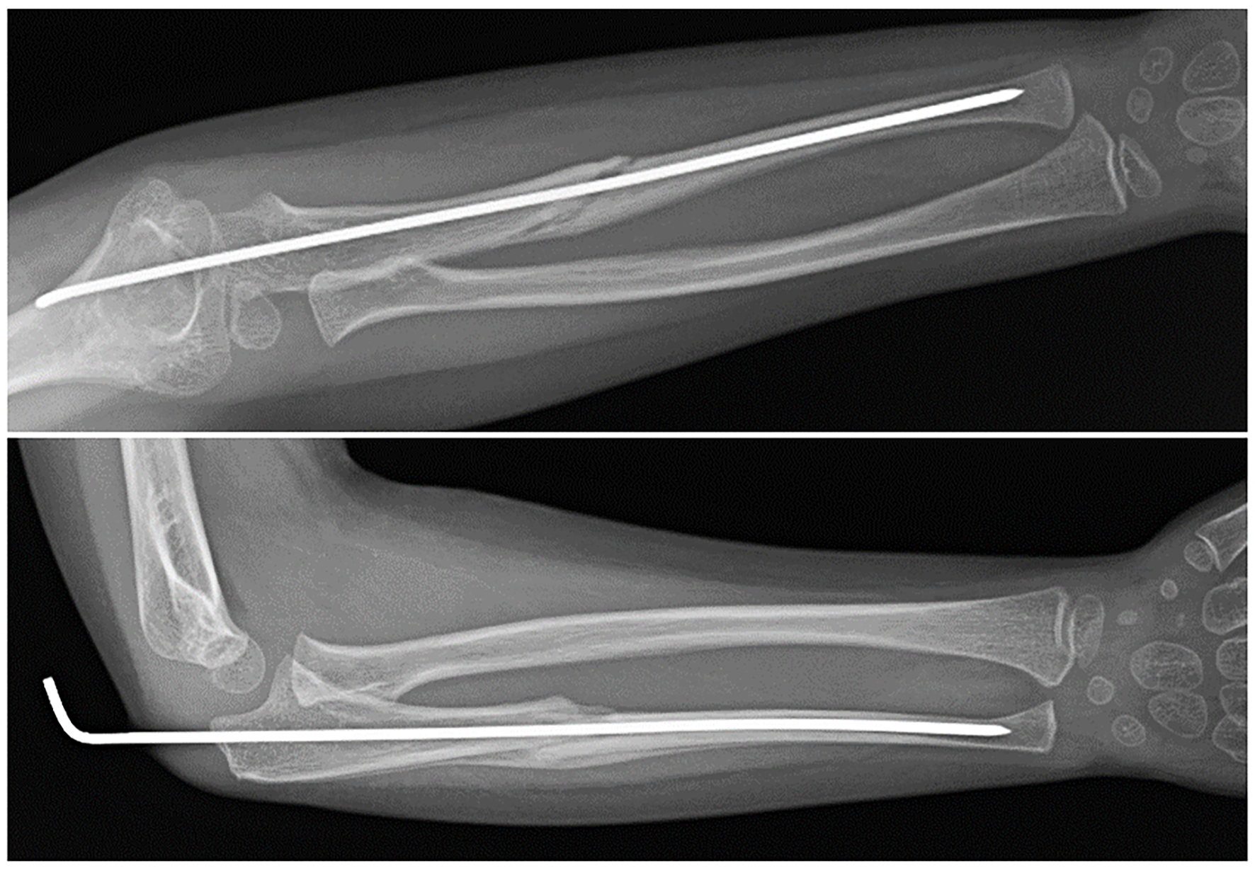
On the same day, the patient underwent surgical treatment of the Monteggia fracture. The surgeon preferred not to use a plate to avoid a delay in fracture healing and to allow the micromotion necessary for callus formation. The operation comprised percutaneous fixation with an elastic intramedullary K-wire of the ulnar fracture and, subsequently, humeroradial joint reduction through a combination of longitudinal traction and manual manipulation. The wire was inserted through the tip of the olecranon and advanced across the fracture site after a reduction is obtained. The reduction of the radial head may be considered stable under fluoroscopic control if the intramedullary fixation of the ulnar fracture maintains its reduced position through flexion, extension, pronation and supination. Alignment of the ulnar injury by K-wire had simultaneously reduced and stabilized the radial head dislocation. No other K-wire was used to maintain the reduction of the radial head. The K-wire can be bent and cut outside the skin to facilitate easy removal. A long arm cast was applied at the end of the operation with the elbow at 90° of flexion and the forearm in supination. Adequate analgesia was provided after surgery, the affected limb was elevated, cryotherapy applied and circulation of the fingers monitored. The girl’s parents were taught how to manage post-operative swelling and pain over the following days. Cast immobilization was used after the surgical treatment until there was radiographic evidence of fracture union.
At the first (5 d) and second (10 d) follow-up, control two-plane radiographs were obtained to assess any possible loss of articular contact between the proximal radius and humeral capitellum and loss of ulnar reduction (> 10 degree increase in the ulnar angle). Healing of the fracture occurred within six weeks after surgery. In children, although the fracture may still be visible at six weeks, adequate healing is represented by a callus on at least three cortices on standard radiographs (Figure 2). At the last follow-up (6 wk), the cast and the intramedullary wire were removed. Dislocation /subluxation, defined as a loss of articular contact between the proximal radius and capitellum, or loss of ulnar reduction, was not apparent in the final X-ray examination. Active range of motion exercises were started on the first day after removal of the cast and K-wire with the aid of a physiotherapist until full elbow range of motion was achieved. No pain, limitations in range of motion or function, or any complications (recurrent dislocation, elbow dysfunction or stiffness, refracture, transient neuropraxias) were observed at the end of the rehabilitative treatment. The patient had excellent results and complete return of elbow motion at short-term clinical follow-up (6 wk after physiotherapy). Complete fracture healing, defined as full return to activities of daily living and sports, occurred four months after the trauma.
If the radial head dislocation or subluxation in Monteggia fractures is not diagnosed and adequately treated, it may lead to chronic elbow disability, pain, progressive valgus deformity, neurologic problems (posterior interosseous nerve palsy), radial head dysplasia and degenerative arthritis, elbow stiffness and loss of motion, particularly supination and pronation[4,7,8]. Foran et al[4], in a retrospective study performed on 94 patients, asserts that 83% of patients are successfully managed with a cast and do not require surgical stabilization. Reduction of the deformity of the ulnar fracture is achieved through a combination of longitudinal traction, elbow rotation and manual manipulation; when the ulnar length has been re-established, the radiocapitellar joint will often reduce spontaneously or as a result of pressure on the radial head. The author asserts that paediatric Monteggia fractures, including in patients with length-unstable ulna fractures, can initially be managed non-operatively, without compromising outcomes or complications; surgery should only be pursued when a conservative approach fails, with the aim of avoiding surgical overtreatment of these injuries. Compared with adults, children with Monteggia fractures have a higher chance of success without surgery for several reasons, including their thicker periosteum (which helps maintain stability of the ulna), less complex ulna fracture patterns, typically lower energy mechanisms, faster healing times and greater remodeling potential[9,10]. Failure or loss of reduction and late instability may occur in up to 20% of cases with this conservative treatment because of the deforming muscular forces and joint disruption[11,12]. Therefore, although most acute Monteggia fractures are treated non-operatively, certain fracture patterns require surgical stabilization[4]. The indications for surgical treatment are complete fracture, unstable and irreducible fractures, open fractures, and fractures with neurovascular compromise[5]. Variations in the treatment of Monteggia fractures can be found among multiple authors. Ring et al[13] affirmed that maintaining ulnar length and anatomic alignment was the key to treating acute Monteggia injuries, because the radial head will remain reduced through healing of the ulnar fracture. Treatment algorithms, based upon the ulnar fracture pattern, have the purpose of minimizing complications and maximizing outcomes through restoration and maintenance of ulnar alignment. The treatment algorithms assert that length-stable fracture patterns with incomplete fracture or plastic deformation of the ulna are treated with closed reduction and casting; length-stable fractures with a complete fracture of the ulna (transverse or short oblique) are treated with intramedullary pin fixation; and length-unstable fractures with comminution of the ulna or a long oblique fracture (fracture line measuring more than twice the cortical diameter) of the ulna are treated with plate fixation[13]. The objective of this algorithm is to avoid loss of reduction and its associated morbidity. Ramski et al[11], in a retrospective study performed on 112 patients, asserts that recurrent instability of the radial head and loss of ulnar fracture reduction only occurred in patients who were not treated according to the ulnar-based algorithm. Failures occurred in patients treated less rigorously than the recommended algorithm, and in particular, failures occurred in complete fractures treated non-operatively with closed reduction and cast immobilization[11]. Furthermore, the authors observed that although the algorithm strategy recommends plate fixation for all long oblique or comminuted fractures, there were no failures of intramedullary pin fixation of long oblique fractures in this clinical study.
Nevertheless, several authors advocate more aggressive treatment with open reduction and plate fixation for unstable fractures[14]. Leonidou et al[14], in a retrospective study of 40 paediatric Monteggia fractures, asserts that although conservative management has been shown to correlate with good results, unstable fractures of the ulna need to be treated with plate fixation to ensure good reduction of the radial head and to avoid the possibility of prolonged immobilization leading to elbow stiffness in children[14]. Moreover, Hetthéssy et al[3] in a retrospective analysis of 23 acute pediatric Monteggia fractures, affirms that if the dislocation of the radial head is accompanied by an unstable fracture of the ulna, a more stable osteosynthesis plate is required.
Nowadays, intramedullary nailing fixation is the main method of fracture stabilization in acute, stable and unstable Monteggia injuries, while plate fixations are widely used in patients with neglected Monteggia fractures[15]. Many authors use a cut-off of fourweeks before considering a Monteggia fracture to be “neglected”. He et al[15], through a retrospective comparison study of 42 patients, showed that the rate of patients that needed open reduction or complex immobilization methods was higher in the neglected group than in the acute group. Therefore, only for early diagnosis of Monteggia injury can we rely upon minimally invasive methods such as intramedullary pin fixation[15-17]. Open reduction combined with ulnar plate fixation is the most common approach to treating missed Monteggia fractures or acute Monteggia fractures in the case of failure of closed radiocapitellar joint reduction[15].
In contrast to short oblique or transverse ulna fractures, long oblique and comminuted fractures are considered “length unstable” and therefore are treated with plate and screw fixation; open reduction and plate fixation are frequently indicated by some orthopaedists because angulation, malalignment and shortening of the ulna often occur after closed reduction and intramedullary wire fixation. In this case report, intramedullary fixation of the ulna allowed a stable reduction and ulnar length and alignment to be achieved as well as radio capitellar reduction despite the fact that the orthopaedic surgeon did not use the plate and screws for ulnar fracture. Therefore, even if not indicated in treatment algorithms, length-unstable Monteggia fractures can be successfully treated with an elastic intramedullary device with the aim of using a minimally invasive technique and enablinga low complication rate.
Treatment of acute paediatric Monteggia fractures is still being debated and there is currently no standard treatment. In fresh Monteggia fractures, the decision to treat the ulna with an intramedullary device or plateismade according tothe surgeon’sown preference with the aim offacilitating the correct alignment of the radiocapitellar joint and avoiding recurrent dislocation. In recent years, the use of intramedullary nails for the management of acute Monteggia fractures in children has gained popularity regardless of the ulnar fracture type. Delayed diagnosis and inadequate treatment of radial head dislocation will cause complications such as elbow pain, decreased joint range of motion, increased valgus deformity and neurologic problems. Flexible nails or wires are excellent devices combining stability and elasticity in children with fractures. Intramedullary fixation of unstable Monteggia fractures results in excellent outcomes, providing reliable reduction and causing fewer complications.
Provenance and peer review: Unsolicited article; Externally peer reviewed
Specialty type: Orthopedics
Country/Territory of origin: Italy
Peer-review report’s scientific quality classification
Grade A (Excellent): 0
Grade B (Very good): B
Grade C (Good): 0
Grade D (Fair): 0
Grade E (Poor): 0
P-Reviewer: Zyoud SH S-Editor: Liu M L-Editor: A P-Editor: Liu M
| 1. | Bruce HE, Harvey JP, Wilson JC Jr. Monteggia fractures. J Bone Joint Surg Am. 1974;56:1563-1576. [PubMed] |
| 2. | Bado JL. The Monteggia lesion. Clin Orthop Relat Res. 1967;50:71-86. [PubMed] |
| 3. | Hetthéssy JR, Sebők B, Vadász A, Kassai T. The Three Step Approach to the management of acute pediatric Monteggia lesions. Injury. 2021;52 Suppl 1:S57-S62. [RCA] [PubMed] [DOI] [Full Text] [Cited by in Crossref: 4] [Cited by in RCA: 5] [Article Influence: 1.3] [Reference Citation Analysis (0)] |
| 4. | Foran I, Upasani VV, Wallace CD, Britt E, Bastrom TP, Bomar JD, Pennock AT. Acute Pediatric Monteggia Fractures: A Conservative Approach to Stabilization. J Pediatr Orthop. 2017;37:e335-e341. [RCA] [PubMed] [DOI] [Full Text] [Cited by in Crossref: 27] [Cited by in RCA: 27] [Article Influence: 3.4] [Reference Citation Analysis (0)] |
| 5. | Poutoglidou F, Metaxiotis D, Kazas C, Alvanos D, Mpeletsiotis A. Flexible intramedullary nailing in the treatment of forearm fractures in children and adolescents, a systematic review. J Orthop. 2020;20:125-130. [RCA] [PubMed] [DOI] [Full Text] [Cited by in Crossref: 13] [Cited by in RCA: 21] [Article Influence: 4.2] [Reference Citation Analysis (0)] |
| 6. | Fernandez FF, Langendörfer M, Wirth T, Eberhardt O. Failures and complications in intramedullary nailing of children's forearm fractures. J Child Orthop. 2010;4:159-167. [RCA] [PubMed] [DOI] [Full Text] [Cited by in Crossref: 46] [Cited by in RCA: 63] [Article Influence: 4.2] [Reference Citation Analysis (0)] |
| 7. | Beutel BG. Monteggia fractures in pediatric and adult populations. Orthopedics. 2012;35:138-144. [RCA] [PubMed] [DOI] [Full Text] [Cited by in Crossref: 35] [Cited by in RCA: 21] [Article Influence: 1.6] [Reference Citation Analysis (0)] |
| 8. | Miller TC, Fishman FG. Management of Monteggia Injuries in the Pediatric Patient. Hand Clin. 2020;36:469-478. [RCA] [PubMed] [DOI] [Full Text] [Cited by in Crossref: 4] [Cited by in RCA: 6] [Article Influence: 1.2] [Reference Citation Analysis (0)] |
| 9. | Rodgers WB, Waters PM, Hall JE. Chronic Monteggia lesions in children. Complications and results of reconstruction. J Bone Joint Surg Am. 1996;78:1322-1329. [RCA] [PubMed] [DOI] [Full Text] [Cited by in Crossref: 127] [Cited by in RCA: 101] [Article Influence: 3.5] [Reference Citation Analysis (0)] |
| 10. | Sessa G, Evola FR, Costarella L. Osteosynthesis systems in fragility fracture. Aging Clin Exp Res. 2011;23:69-70. [PubMed] |
| 11. | Ramski DE, Hennrikus WP, Bae DS, Baldwin KD, Patel NM, Waters PM, Flynn JM. Pediatric monteggia fractures: a multicenter examination of treatment strategy and early clinical and radiographic results. J Pediatr Orthop. 2015;35:115-120. [RCA] [PubMed] [DOI] [Full Text] [Cited by in Crossref: 49] [Cited by in RCA: 37] [Article Influence: 3.7] [Reference Citation Analysis (0)] |
| 12. | Peshin C, Ratra R, Juyal AK. Step-cut osteotomy in neglected Monteggia fracture dislocation in pediatric and adolescent patients: A retrospective study. J Orthop Surg (Hong Kong). 2020;28:2309499020964082. [RCA] [PubMed] [DOI] [Full Text] [Cited by in Crossref: 2] [Cited by in RCA: 3] [Article Influence: 0.8] [Reference Citation Analysis (0)] |
| 13. | Ring D, Jupiter JB, Waters PM. Monteggia fractures in children and adults. J Am Acad Orthop Surg. 1998;6:215-224. [RCA] [PubMed] [DOI] [Full Text] [Cited by in Crossref: 128] [Cited by in RCA: 86] [Article Influence: 3.2] [Reference Citation Analysis (0)] |
| 14. | Leonidou A, Pagkalos J, Lepetsos P, Antonis K, Flieger I, Tsiridis E, Leonidou O. Pediatric Monteggia fractures: a single-center study of the management of 40 patients. J Pediatr Orthop. 2012;32:352-356. [RCA] [PubMed] [DOI] [Full Text] [Cited by in Crossref: 22] [Cited by in RCA: 21] [Article Influence: 1.6] [Reference Citation Analysis (0)] |
| 15. | He JP, Hao Y, Shao JF. Comparison of treatment methods for pediatric Monteggia fracture: Met vs missed radial head dislocation. Medicine (Baltimore). 2019;98:e13942. [RCA] [PubMed] [DOI] [Full Text] [Full Text (PDF)] [Cited by in Crossref: 11] [Cited by in RCA: 10] [Article Influence: 1.7] [Reference Citation Analysis (0)] |
| 16. | Kruppa C, Bunge P, Schildhauer TA, Dudda M. Low complication rate of elastic stable intramedullary nailing (ESIN) of pediatric forearm fractures: A retrospective study of 202 cases. Medicine (Baltimore). 2017;96:e6669. [RCA] [PubMed] [DOI] [Full Text] [Full Text (PDF)] [Cited by in Crossref: 30] [Cited by in RCA: 40] [Article Influence: 5.0] [Reference Citation Analysis (0)] |
| 17. | Yuan Z, Xu H, Li Y, Li J, Liu Y, Canavese F. Functional and radiological outcome in patients with acute Monteggia fracture treated surgically: a comparison between closed reduction and external fixation versus closed reduction and elastic stable intramedullary nailing. J Pediatr Orthop B. 2020;29:438-444. [RCA] [PubMed] [DOI] [Full Text] [Cited by in Crossref: 5] [Cited by in RCA: 5] [Article Influence: 1.0] [Reference Citation Analysis (0)] |


