Copyright
©The Author(s) 2018.
World J Orthop. Sep 18, 2018; 9(9): 138-148
Published online Sep 18, 2018. doi: 10.5312/wjo.v9.i9.138
Published online Sep 18, 2018. doi: 10.5312/wjo.v9.i9.138
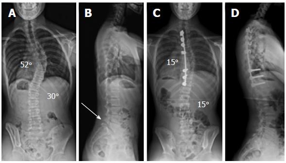
Figure 1 Patient with adolescent idiopathic scoliosis (patient 13; Table 4).
A: Preoperative postero-anterior spinal radiograph shows a primary thoracic and compensatory lumbar AIS; B: Preoperative lateral spinal radiograph shows thoracic hypokyphosis and isthmic spondylolysis at L5 (white arrow) with associated grade 1 lumbosacral spondylolisthesis; C: Postero-anterior spinal radiograph shows very satisfactory scoliosis correction across both curves and a globally balanced spine following a selective posterior thoracic fusion; D: Lateral spinal radiograph shows restoration of thoracic kyphosis and a normal sagittal balance of the spine with no change in the grade 1 lumbosacral spondylolisthesis. AIS: Adolescent idiopathic scoliosis.
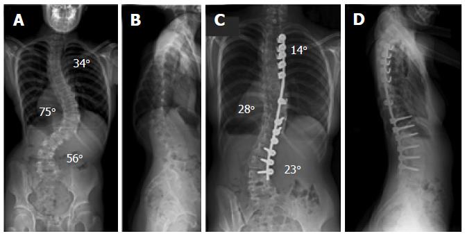
Figure 2 Patient with neuromuscular kyphoscoliosis.
A: Preoperative postero-anterior spinal radiograph shows a triple thoracic and lumbar scoliosis; B: Preoperative lateral spinal radiograph shows a thoracolumbar rotatory kyphosis producing positive global sagittal balance of the spine; C: Postero-anterior spinal radiograph shows excellent scoliosis correction and a balanced spine in the coronal plane; D: Lateral spinal radiograph shows restoration of thoracic kyphosis and a normal sagittal balance of the spine.
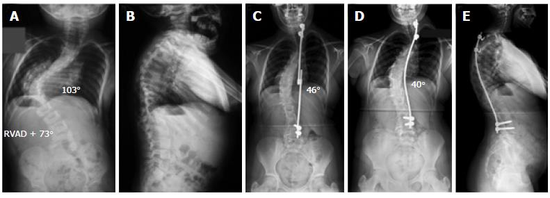
Figure 3 Patient with a severe infantile idiopathic thoracic scoliosis.
A: Initial postero-anterior spinal radiograph at age 2 years shows a very severe thoracic scoliosis; B: Initial lateral spinal radiograph at age 2 years shows increased thoracic kyphosis. The patient was treated with placement of a concave growing rod construct followed by 21 consecutive lengthening procedures; C: Postero-anterior spinal radiograph at age 13 years when she underwent the definitive posterior spinal fusion with the use of a single concave rod construct; D: Postero-anterior spinal radiograph at latest follow-up (end of spinal growth) shows no evidence of recurrence of the deformity and no crankshaft effect with a globally balanced spine; E: Lateral spinal radiograph shows good global balance in the sagittal plane at skeletal maturity.
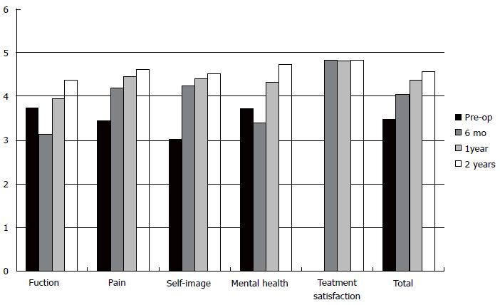
Figure 4 Scoliosis Research Society (SRS-22) in our adolescent idiopathic scoliosis patient population.
Mean scores are presented pre-operatively, at 6-mo, one-year, and two-years post-operatively including postoperative patient satisfaction. There was statistically significant improvement of individual domain scores with regards to function, pain, self-image, mental health, and total score between preoperative and 2-year values (P < 0.001).
- Citation: Tsirikos AI, Loughenbury PR. Single rod instrumentation in patients with scoliosis and co-morbidities: Indications and outcomes. World J Orthop 2018; 9(9): 138-148
- URL: https://www.wjgnet.com/2218-5836/full/v9/i9/138.htm
- DOI: https://dx.doi.org/10.5312/wjo.v9.i9.138


