Copyright
©The Author(s) 2017.
World J Orthop. Mar 18, 2017; 8(3): 271-277
Published online Mar 18, 2017. doi: 10.5312/wjo.v8.i3.271
Published online Mar 18, 2017. doi: 10.5312/wjo.v8.i3.271
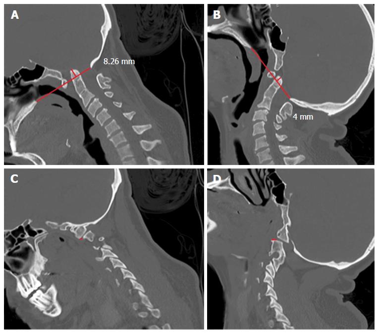
Figure 1 A 18-year-old woman with basilar invagination and tonsillar herniation of 7 mm.
She also had atlas assimilation. A: Sagittal computed tomography (CT) scan in flexion shows the tip of the odontoid 8.26 mm above the Chamberlain’s line; B: Sagittal CT scan in extended position shows the tip of the odontoid 4 mm above the Chamberlain’s line; C: Sagittal CT scan showing anterior dislocation of the facet joint of C1 over C2 facetary of 2 mm; D: Sagittal CT scan showing posterior dislocation of the C1 facet joint over the facet of C2 of 3 mm, ranging 5 mm in dynamic exam. This patient underwent a craniocervical fusion concomitant to the posterior fossa decompression.
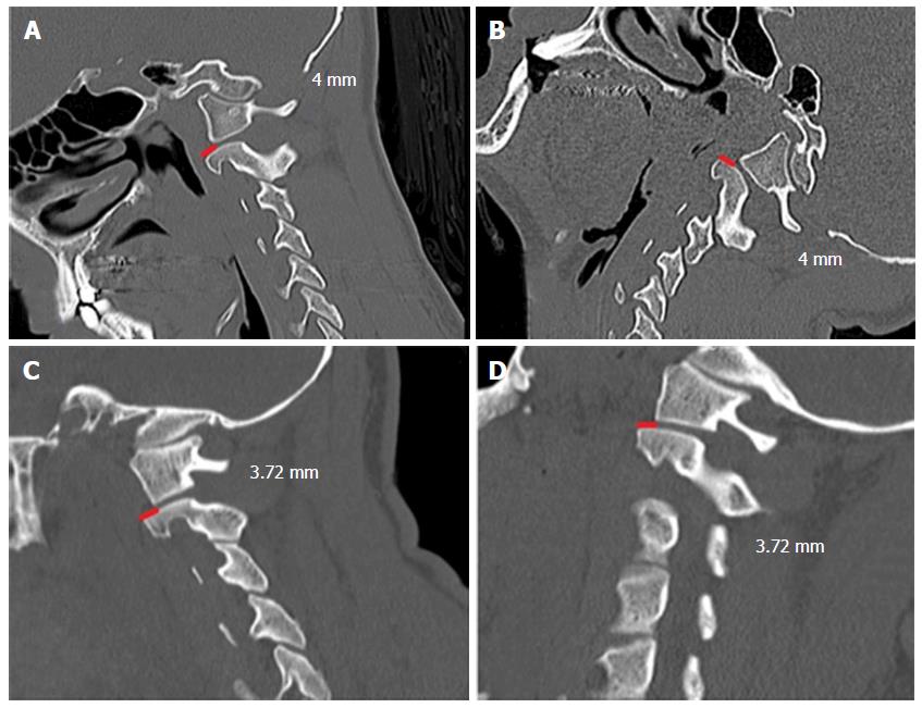
Figure 2 Dynamic sagittal computed tomography scan of 15-year-old boy, which had the diagnosis of a tonsilar herniation of 9.
4 mm and basilar invagination. In both A and B positions, the facet dislocation of the C1 lateral mass posteriorly to the superior C2 joints was maintained (4 mm). We opted to perform only posterior fossa decompression without craniovertebral junction instrumentation, obtaining a good clinical improvement; C and D: Dynamic sagittal computed tomography scan imaging - a forty-six-year-old man, with tonsilar herniation of 7.44 mm. In both positions the facet dislocation of the C1 lateral mass over the C2 superior facet joint was 3.72 mm. We also performed only a posterior fossa decompression without fusion in this patient, with a good clinical outcome.
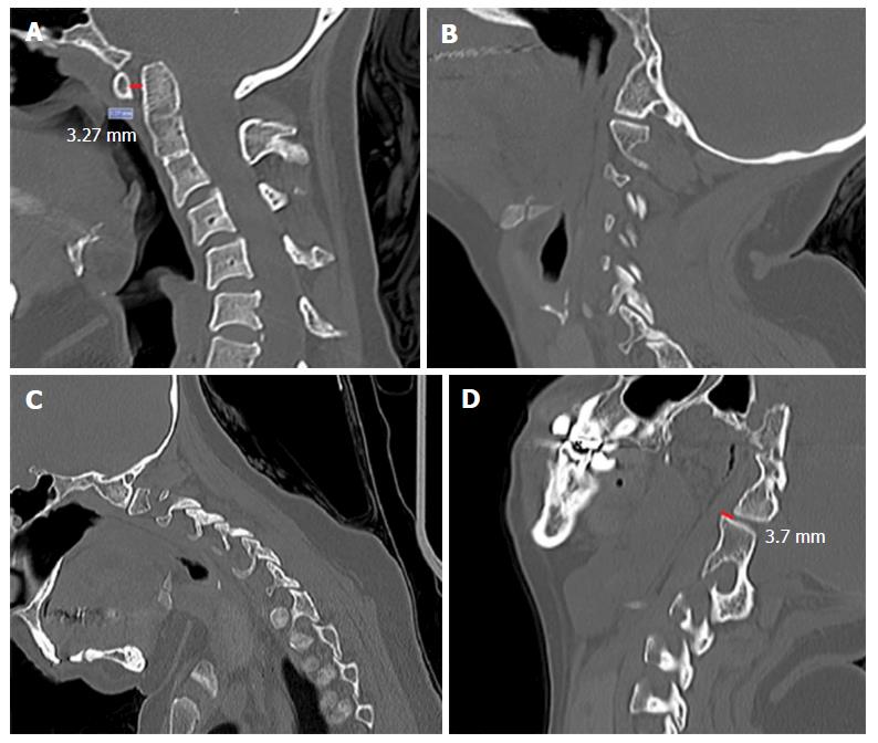
Figure 3 Forty-year-old woman with a tonsilar herniation of 5 mm and basilar invagination.
Noted that she also had atlas assimilation and a congenital C23 fusion A, B and C: Pre-operative dynamic imaging, showing a atlanto-dental interval of 3.27 mm, but no signs of facet dislocation in flexion or in extension (B and C); D: Sagittal computed tomography scan obtained after some months after posterior fossa decompression showing an evident facet joints dislocation (the assimilated lateral mass of C1 was dislocated posteriorly over the superior facet joint of C2). She had some symptoms of dizziness and cervical pain when flexing the neck and an occipto-cervical fixation was proposed but the patient declined surgical treatment because she was not doing well with depression and mood disorders.
- Citation: da Silva OT, Ghizoni E, Tedeschi H, Joaquim AF. Role of dynamic computed tomography scans in patients with congenital craniovertebral junction malformations. World J Orthop 2017; 8(3): 271-277
- URL: https://www.wjgnet.com/2218-5836/full/v8/i3/271.htm
- DOI: https://dx.doi.org/10.5312/wjo.v8.i3.271


