Copyright
©The Author(s) 2017.
World J Orthop. Mar 18, 2017; 8(3): 271-277
Published online Mar 18, 2017. doi: 10.5312/wjo.v8.i3.271
Published online Mar 18, 2017. doi: 10.5312/wjo.v8.i3.271
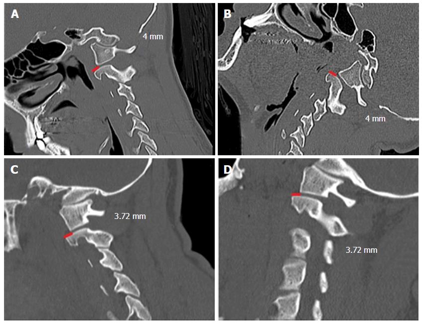
Figure 2 Dynamic sagittal computed tomography scan of 15-year-old boy, which had the diagnosis of a tonsilar herniation of 9.
4 mm and basilar invagination. In both A and B positions, the facet dislocation of the C1 lateral mass posteriorly to the superior C2 joints was maintained (4 mm). We opted to perform only posterior fossa decompression without craniovertebral junction instrumentation, obtaining a good clinical improvement; C and D: Dynamic sagittal computed tomography scan imaging - a forty-six-year-old man, with tonsilar herniation of 7.44 mm. In both positions the facet dislocation of the C1 lateral mass over the C2 superior facet joint was 3.72 mm. We also performed only a posterior fossa decompression without fusion in this patient, with a good clinical outcome.
- Citation: da Silva OT, Ghizoni E, Tedeschi H, Joaquim AF. Role of dynamic computed tomography scans in patients with congenital craniovertebral junction malformations. World J Orthop 2017; 8(3): 271-277
- URL: https://www.wjgnet.com/2218-5836/full/v8/i3/271.htm
- DOI: https://dx.doi.org/10.5312/wjo.v8.i3.271


